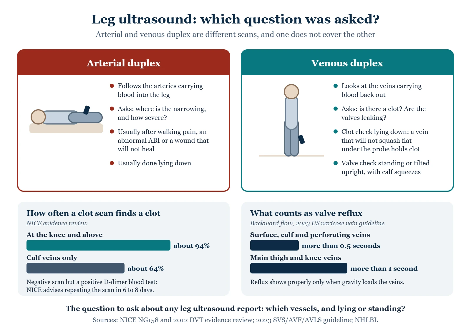
“I had an ultrasound of my leg” does not say which question was asked. Arterial and venous duplex are different studies, looking at different vessels, for different problems — and assuming one covered the other is a common and consequential mistake.
The two studies
| Arterial duplex | Venous duplex | |
|---|---|---|
| Looks at | Arteries carrying blood into the leg | Veins carrying blood back out |
| Main questions | Where is the narrowing, and how severe? | Is there a clot? Are the valves leaking? |
| Typical trigger | Leg pain on walking, an abnormal ABI, a non-healing wound | One swollen or painful leg, varicose veins, skin changes, a planned vein procedure |
| Position | Usually lying down | Lying down to look for clot; standing or tilted upright to test valve reflux |
| What the sonographer does | Follows arteries down the leg, measuring flow speeds | Presses the probe to see whether veins compress, and uses calf squeezes to test valve closure |
The reflux test needs you upright
Valve leakage only shows properly when gravity is loading the veins, which is why the scan is done standing when that is safe. A venous study done entirely lying flat can find a clot but may miss reflux. If you are being assessed for varicose veins or venous insufficiency and the whole scan was done lying down, it is reasonable to ask whether reflux was tested — it changes what the report can tell you.
Reflux has a formal definition. The 2023 US varicose vein guideline counts blood running backward for more than half a second in the surface, calf and perforating veins, or for more than one second in the main thigh and knee veins. A report that gives reflux “in seconds” is measuring exactly that.
Ultrasound for a suspected clot
Compression ultrasound is the main test for a deep vein thrombosis: if a vein does not squash flat under the probe, there is clot in it. It is very good above the knee and less so in the calf. In a NICE evidence review it found about 94% of clots at the knee and above but only about 64% of clots confined to the calf veins, which is why NICE advises repeating the scan six to eight days later when the first scan is negative but the D-dimer blood test is positive. When a clot is likely, NICE advises scanning within four hours. How DVT is diagnosed →
Ultrasound for the arteries
An arterial duplex usually follows symptoms that fit, or an abnormal ankle-brachial index, and maps where the narrowing is and how severe. In diabetes and kidney disease, calcified arteries can make the ABI read falsely normal or high, so toe pressures and the toe-brachial index (0.70 or less is abnormal) add information the cuff at the ankle cannot. The ABI and TBI →
What the test is like
Gel on the skin, a probe moved over the leg, some pressing. It makes swooshing and pulsing sounds as flow is sampled. It is not painful, though pressing over a tender calf can be uncomfortable, and standing for the reflux portion can be tiring. There is no radiation and no injection. Wear something that lets the whole leg be exposed. Scans vary in length with how much of the leg is examined, and the department will tell you how long to allow.
No fasting is needed for leg studies. For abdominal or aortic imaging you may be asked to fast — the department will say.
When ultrasound is not enough
- CT angiography (CTA) — a detailed map of the arteries using contrast dye. Fast and widely available; involves radiation, and kidney function affects the decision.
- MR angiography (MRA) — no radiation, different contrast considerations, less suitable with some implants.
- Catheter angiography — the most detailed, done in a procedure suite, usually at the time treatment is planned or performed.
Heavy calcification in the vessel wall — common in diabetes and kidney disease — can limit what both ultrasound and CT can show, which is one reason more than one test is sometimes needed.
Making sense of your report
Reports are written for clinicians, and the language is not always intuitive. Terms worth knowing: stenosis is narrowing, usually with a percentage; occlusion means completely blocked; reflux means blood flowing backward through a leaky valve, with a duration in seconds; patent means open; non-compressible in a venous report suggests clot, while in an arterial or ABI context it means the vessel is too stiff to squeeze. Monophasic, biphasic and triphasic describe the shape of the arterial waveform, with triphasic being the normal pattern.
If a report has been handed to you without an explanation of what it means for you, asking for one is entirely reasonable.
From the clinic
The question I ask about every leg ultrasound report is simple: which vessels, and lying or standing? A “normal leg ultrasound” that turns out to have been a quick clot scan done lying flat says nothing about the arteries or the valves, and I have met people who were told their circulation was fine on the strength of one.
Common questions
I had an ultrasound and it was normal. Are my arteries fine?
Only if it was an arterial study. A venous duplex looking for a clot says nothing about your arteries. Worth checking which one you had — the report title usually says, and if you are unsure it is a quick question for the ordering clinician. Even on the arterial side, a normal resting test does not always settle it: up to 30% of people with symptoms and a normal resting ABI turn out to be abnormal after walking.
Does ultrasound rule out a clot completely?
Compression ultrasound is very good for clots in the larger leg veins. Clots confined to small calf veins are harder to see: in one NICE evidence review, ultrasound found about 94% of clots at the knee and above but about 64% of calf-only clots. That is why a repeat scan six to eight days later is advised when the first scan is negative but the D-dimer is positive.
Why do I need a CT if I already had an ultrasound?
Usually because a procedure is being planned and the team needs a complete road map — including segments ultrasound cannot see well, such as vessels deep in the pelvis or abdomen.
Is the contrast dye risky?
It carries some risk, mainly relating to kidney function and to allergy. That is exactly what the pre-test questions and blood tests are for, and it is why kidney function comes up when these scans are arranged. Tell the department about previous reactions and about kidney disease.
Why did they make me stand for the vein scan?
Because leaky valves only show their leak when gravity is pulling blood down the leg. Standing lets the sonographer time how long blood runs backward after a squeeze, which is how reflux is measured and graded.
Where to go next
Sources
- National Heart, Lung, and Blood Institute. PAD — Diagnosis.
- NHS. Deep vein thrombosis (DVT).
- Society for Vascular Surgery. Chronic Venous Insufficiency.
- Centers for Disease Control and Prevention. About Venous Thromboembolism.
- NICE. Venous thromboembolic diseases: diagnosis, management and thrombophilia testing (NG158).
- NICE (NCBI Bookshelf). 2012 evidence review: diagnosis of deep vein thrombosis.
- Society for Vascular Surgery, American Venous Forum and American Vein and Lymphatic Society. 2023 clinical practice guidelines for varicose veins of the lower extremities, part II.
- StatPearls. Venous insufficiency.
- Gornik HL, et al. 2024 ACC/AHA multisociety guideline for the management of lower extremity peripheral artery disease. Circulation 2024.
- American Family Physician. Peripheral artery disease: rapid evidence review (2019).
Written by Tom Biernacki, DPM, FACFAS. Published 20 September 2026. Updated 15 October 2026. General education, not medical advice.