A deep vein thrombosis (DVT) is a blood clot in one of the deep veins, usually in the leg. It matters for two reasons: a piece can break off and travel to the lungs, and about half of DVTs cause no symptoms at all. No symptom list can rule one out — but a few hours of testing usually can.
Call 911 now if you have any of these
- Difficulty breathing that came on suddenly
- Chest pain or discomfort, usually worse with a deep breath or coughing
- Coughing up blood
- A faster than normal or irregular heartbeat
- Lightheadedness, fainting, or very low blood pressure
These can be signs of a pulmonary embolism — a clot that has traveled to the lungs. This is an emergency. Outside the United States, use your local emergency number.
Be seen today if you have these
Swelling, pain or tenderness, warmth, or redness or discoloration in one leg — particularly when the other leg is normal, and particularly if it developed over hours to days. You need an assessment and usually an ultrasound, today rather than at the next available appointment.
How common it is
The CDC estimates that up to 900,000 Americans develop a venous blood clot — a DVT or a pulmonary embolism — each year, and that 60,000 to 100,000 die. For about 1 in 4 people with a pulmonary embolism, sudden death is the first symptom. More than a third of these clots follow a recent hospital stay, up to 70% of hospital-related clots are preventable, and after surgery 60% of clots appear once the person has gone home, up to 90 days later. That is why clot prevention is part of hospital care, and why the weeks after discharge matter as much as the stay itself.
Symptoms
When a DVT does cause symptoms, they are usually in one leg — most often the calf — and include:
- Swelling, sometimes of the whole leg, sometimes just the calf or ankle
- Pain, aching or tenderness, often described as a cramp or a pulled muscle that does not settle
- Warmth over the affected area
- Red, darker or bluish skin, which can be harder to see on brown or black skin
- Surface veins that look fuller than on the other leg
The problem is that every one of those also fits a strained muscle, a burst Baker’s cyst behind the knee, a skin infection, or a flare of long-standing vein disease. The overlap is the reason for the testing pathway below. Calf pain: muscle, clot or artery? →
Why timing is the whole story
A leg clot is treatable. Anticoagulation stops it growing and lets the body break it down, and it substantially reduces the chance of a piece traveling to the lungs. The reason urgency matters is not that the leg will be lost — it is that the lung complication is the dangerous one, and the window to prevent it is before it happens.
There is a second, slower reason. A clot damages the vein valves it sits against. That damage drives post-thrombotic syndrome — chronic swelling, aching and skin change in that leg, sometimes years later. More on post-thrombotic syndrome →
What raises the risk
- Recent surgery, particularly hip, knee or abdominal, and recent major trauma; the risk is highest in the first three months
- Prolonged immobility — hospital admission, a plaster cast or boot, a long flight or drive of more than about four hours
- Cancer and some cancer treatments; about 1 in 5 clots is linked to cancer
- Pregnancy and the three months after delivery, when the risk is about five times higher
- Estrogen-containing contraception and hormone therapy, with the risk highest in the first few months of use
- A previous clot, or a family history of clots
- Inherited or acquired clotting disorders, such as factor V Leiden, which is carried by 3 to 8% of people of European ancestry
- Obesity, smoking, and older age; the risk roughly doubles with each decade after 40
- Central venous catheters
Risk factors raise the probability; their absence does not remove it. Plenty of clots happen in people with none of these.
Why we will not give you a checklist that rules it out
Because none exists. Around half of deep vein clots produce no symptoms. Of those that do, the symptoms — swelling, ache, warmth, redness — overlap almost completely with a pulled muscle, a ruptured Baker’s cyst, cellulitis and a flare of chronic vein disease. Clinicians do not exclude DVT by examination either; they use a structured risk assessment, a blood test and an ultrasound. That is the honest answer, and it is why the advice here is simply to be seen.
How it is diagnosed
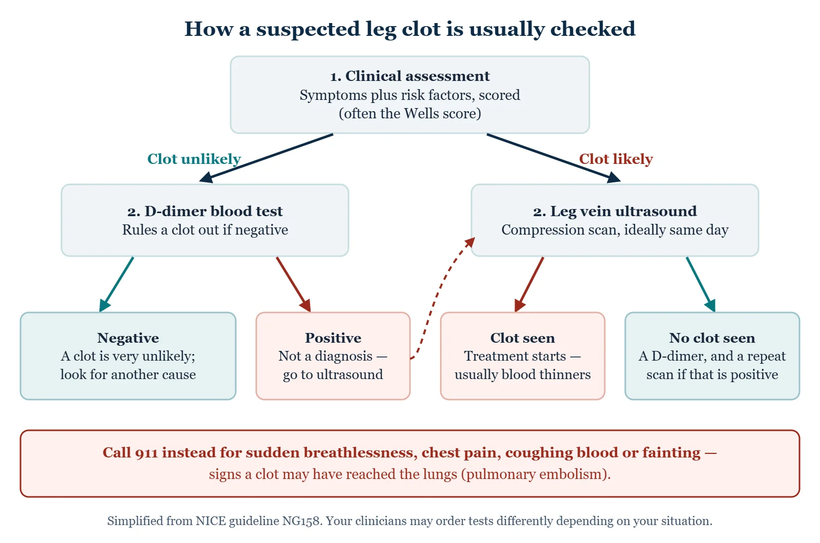
- A structured clinical assessment combining symptoms and risk factors into a probability, most often with a tool called the Wells score. It gives a point for each of nine features, such as active cancer, recent surgery or bed rest, a calf more than 3 cm larger than the other, or a previous DVT, and subtracts two if another diagnosis is as likely. A score of 2 or more means a clot is “likely”.
- A D-dimer blood test in lower-probability situations. A negative result in that setting effectively rules a clot out; a positive one is non-specific — it also rises after surgery, in pregnancy, with infection and with age — and leads to imaging. Over the age of 50, some services adjust the cut-off for age.
- Compression ultrasound of the leg veins — the main test. If the vein does not compress under the probe, there is clot in it. In the UK, NICE advises that when a clot is likely the scan should happen within four hours; if it cannot, a D-dimer is taken, a blood thinner started, and the scan done within 24 hours. Ultrasound is very good at finding clots at the knee and above but misses more in the calf (in one analysis it found 94% of the former and 64% of the latter). If the first scan is negative but the D-dimer is positive, NICE advises repeating the scan six to eight days later.
- Imaging of the chest — usually CT pulmonary angiography — if pulmonary embolism is suspected.
Note that this is a venous ultrasound. It is a different study from an arterial ultrasound, and “I had a leg ultrasound” does not tell you which question was asked. More on the two ultrasounds →
Treatment
Anticoagulation — blood-thinning medicine — is the mainstay. It does not dissolve the clot directly; it stops it growing and stops new clot forming while the body breaks down what is there.
| Option | What it is | When it tends to be used |
|---|---|---|
| Direct oral anticoagulants (DOACs) | Tablets such as apixaban, rivaroxaban, dabigatran and edoxaban | First choice for most people; no routine blood-level monitoring |
| Low-molecular-weight heparin | Injections under the skin | Pregnancy, some cancers, and as a lead-in before some tablets |
| Warfarin | Tablet needing regular INR blood tests | Certain heart valves, severe kidney disease, antiphospholipid syndrome, cost |
| Clot-dissolving or clot-removal procedures | Catheter-directed treatment in a specialist unit | Selected large clots, such as extensive thigh-and-pelvis clots threatening the leg |
| Vena cava filter | A small device placed in the main abdominal vein | Only when blood thinners cannot be used |
Treatment usually lasts at least three months. After a clot with a major temporary trigger, such as surgery, the CHEST guideline recommends stopping there. After a clot with no trigger, or with an ongoing risk factor, it recommends offering open-ended treatment, often at a reduced dose (for example apixaban 2.5 mg twice daily or rivaroxaban 10 mg daily), reviewed every year. Most people with an uncomplicated leg clot are treated at home rather than admitted, as the American Society of Hematology suggests; hospital is for very large clots, high bleeding risk, other serious illness, or too little support at home.
These decisions turn on details of your own history and bleeding risk. This page is a map, not a plan.
A clot only in the calf
Clots confined to the smaller veins of the calf, below the knee, are sometimes managed differently. For a low-risk calf-only clot with mild symptoms, the CHEST guideline suggests repeating the ultrasound weekly for two weeks instead of starting a blood thinner, and treating only if the clot extends; severe symptoms or risk factors for spreading favor treatment from the start. About 10 to 15% of untreated calf clots spread upward, and the guideline notes that newer data more clearly favor treatment, which cut three-month recurrence from 9.1% to 2.9%.
After a DVT
- Walking is encouraged once treatment has started. Bed rest is no longer advised for a leg clot.
- Swelling and aching usually settle over weeks, sometimes months. Raising the leg when resting helps.
- Compression stockings are no longer recommended routinely to prevent post-thrombotic syndrome. In the placebo-controlled SOX trial, 14.2% of people in real stockings developed it against 12.7% in placebo stockings, and NICE, CHEST and the American Society of Hematology all advise against routine use for that purpose. They can still ease swelling and aching, so if they make the leg more comfortable they are reasonable to wear.
- Know the bleeding warning signs on blood thinners — black or bloody stools, blood in the urine, vomiting blood, a severe headache, or bleeding that will not stop — and get help promptly if they happen.
- Tell every clinician and dentist that you take a blood thinner before any procedure.
- Clots can come back. The CDC estimates that a third of people with a venous clot have another within 10 years. In the CHEST guideline’s figures, five years after stopping treatment the risk is about 3% when surgery caused the clot, 15% after a non-surgical trigger and 30% when there was no trigger, which is why the length of treatment depends so much on the cause.
Lowering the risk
- In hospital or after surgery: ask what your clot-prevention plan is. It may include injections, tablets, stockings or calf pumps, and getting up and walking as soon as you safely can.
- On journeys over about four hours: walk the aisle, flex your ankles and calves, and keep hydrated. If you have a previous clot or other strong risk factors, ask about properly fitted below-knee stockings. Aspirin taken only for the trip is not advised for most travelers. Long flights and long drives →
- Before starting estrogen-containing contraception or hormone therapy: mention any personal or family history of clots.
- Every day: stay active, do not smoke, and keep to a healthy weight.
From the clinic
In foot and ankle work, a cast or walking boot after surgery or an injury is one of the classic setups for a leg clot: the calf pump stops working, and the leg sits still for weeks. A calf that becomes newly swollen, tight or painful inside a boot is not “just the boot”. Call the team that put it on that day — do not wait for the next follow-up appointment, and do not loosen the straps and hope.
Common questions
Can I tell a DVT from a pulled muscle?
Not reliably, and neither can a clinician without testing. A muscle injury usually has a story — a specific movement, a moment it happened. A clot often does not. But plenty of clots are blamed on an imagined strain, which is exactly how they get missed. One newly swollen or painful calf with no clear injury is a reason to be checked.
Should I massage my calf or stretch it out?
No. If a clot is a possibility, leave the leg alone and get it assessed rather than manipulating it. Get the ultrasound first and the physical therapy afterwards if it turns out to be a muscle.
I have no risk factors. Can I relax?
No. A substantial share of clots occur in people with no identifiable risk factor. The absence of risk factors lowers the probability; it does not make the ultrasound unnecessary when the leg looks like this.
Can a DVT go away on its own?
Some small clots do dissolve without treatment, but there is no way to know in advance which ones, and an untreated clot can grow or travel to the lungs. That is why a confirmed DVT in the main leg veins is treated, and why a calf-only clot that is not treated is watched with repeat ultrasound.
How long will I be on blood thinners?
Usually at least three months. After a clot with a clear temporary trigger, such as surgery, treatment often stops there. After a clot with no obvious cause, or a repeat clot, longer or indefinite treatment is often discussed, weighing the risk of another clot against the risk of bleeding.
Can I walk with a DVT?
Yes — once treatment has started, walking is encouraged rather than discouraged. It helps the swelling and the calf pump, and staying in bed does not make the clot safer.
Does a clot mean I will always have a swollen leg?
Not necessarily, but post-thrombotic syndrome is a real possibility — persistent swelling, aching and skin change in that leg, developing over months to years. It affects between a third and a half of people after a DVT, and is severe in up to 5%. Taking the anticoagulation as prescribed, staying active and following the advice you are given about compression all help.
Can I fly after a DVT?
Usually yes, once treatment is established. The NHS advises waiting at least two weeks after starting blood thinners before a flight or long journey, and the British Thoracic Society suggests two weeks after diagnosis. Beyond that the timing and precautions are individual, so ask the team treating you rather than working from general travel advice.
Where to go next
Sources
- CDC. Data and statistics on venous thromboembolism.
- CDC. About venous thromboembolism (blood clots).
- CDC. Healthcare-associated venous thromboembolism.
- CDC. Blood clots and travel.
- CDC Yellow Book. Deep vein thrombosis and pulmonary embolism.
- National Heart, Lung, and Blood Institute. Venous thromboembolism: causes and risk factors.
- National Heart, Lung, and Blood Institute. Venous thromboembolism in women.
- National Heart, Lung, and Blood Institute. Recovery after venous thromboembolism.
- NHS. Deep vein thrombosis (DVT).
- NICE. Venous thromboembolic diseases: diagnosis, management and thrombophilia testing (NG158).
- NICE (NCBI Bookshelf). 2012 evidence review: diagnosis of deep vein thrombosis.
- CHEST. Antithrombotic therapy for VTE disease: second update of the CHEST guideline (2021).
- American Society of Hematology. 2020 guidelines for management of venous thromboembolism: treatment of DVT and PE.
- Cochrane. Compression therapy for prevention of post-thrombotic syndrome.
- StatPearls. Post-thrombotic syndrome.
- MedlinePlus Genetics. Factor V Leiden thrombophilia.
- Mayo Clinic. Deep vein thrombosis: diagnosis and treatment.
- Cleveland Clinic. Pulmonary embolism.
Written by Tom Biernacki, DPM, FACFAS. Published 20 September 2026. Updated 15 October 2026. General education, not medical advice. This page has not yet been through the independent clinical review described on our medical review page.