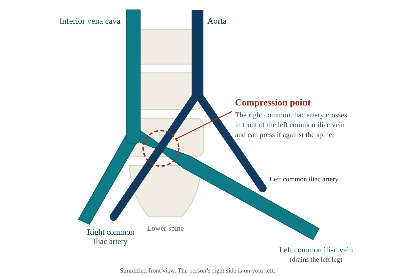
May-Thurner syndrome is a squeeze deep in the pelvis: the artery that supplies the right leg crosses over the main vein draining the left leg and presses it against the lower spine. About one person in five has some degree of this compression, and most never know it. It matters when the narrowing causes a clot or lasting swelling in the left leg — and then it changes how that clot is treated.
If a leg has newly become swollen or painful
Be seen today — emergency department, urgent care, or same-day primary care, wherever can arrange an ultrasound now. That applies to swelling, pain, warmth or a change in skin color in one leg over hours to days, including a leg that already has a vein stent.
Call 911 for:
- Sudden breathlessness, any chest pain or discomfort, coughing up blood, a racing or irregular heartbeat, lightheadedness or fainting. These can mean a clot has reached the lungs.
- A leg that becomes massively swollen, bluish and painful, sometimes within hours. This is rare, and it is an emergency.
Iliac vein compression: what is being squeezed
Blood from each leg returns toward the heart through a large vein in the pelvis, the common iliac vein. On its way up, the left one passes behind the right common iliac artery. In May-Thurner syndrome — also called iliac vein compression syndrome or Cockett syndrome — that artery presses the vein against the lumbar spine and narrows it.
Over years, the artery’s pulsing can irritate the lining of the vein until fibrous bands, called spurs, form across the channel. This scarring is not expected to reverse, which is why treatment, when needed, usually means holding the vein open.
Because the squeezed vein drains the left leg, that is where symptoms usually appear. Right-sided variants have been described but are much less common.
Who it affects
Women are diagnosed about twice as often as men, most often between the ages of 20 and 50. The classic story is a younger woman whose left leg swells after surgery, a long spell of immobility, pregnancy or childbirth, or starting estrogen-containing contraception — each of which adds clot risk to the narrowed vein. The Society for Vascular Surgery also lists a previous clot, obesity, prolonged sitting or standing, and a family history of the condition as risk factors.
The syndrome is estimated to cause 2% to 5% of all deep vein clots, but imaging studies of people with a left-leg clot have found it in 22% to 76% of them. It is thought to be underdiagnosed.
May-Thurner syndrome symptoms
When the compression does cause trouble, it shows up in one of these ways, usually in the left leg.
| Pattern | What it can look like | Urgency |
|---|---|---|
| A clot (DVT) starting high in the leg | Swelling of the left leg, often all of it, with pain, heaviness, warmth or a change in skin color over hours to days | Today |
| Long-standing swelling | A heavy, aching or tight leg that builds through the day and eases with rest or elevation | Routine |
| Vein and skin changes | Varicose veins, darkened skin, and in time an open sore (ulcer) | Routine; same day or next day for an open wound |
| Venous claudication | Leg pain that comes on with walking | Routine |
| Pelvic pain, in some women | Chronic pelvic pain from congested pelvic veins | Routine |
May-Thurner clots tend to start high, in the large veins of the pelvis and thigh, and extensive clots like these often leave post-thrombotic syndrome behind — lasting swelling, aching and skin change in that leg.
Slower symptoms overlap with chronic venous insufficiency, which the compression can itself cause. For a swollen leg with no explanation yet, start with one swollen leg, where the list of possible causes is much longer.
Why most people with the anatomy have no symptoms
The crossing is usually harmless. Where the vein is narrowed, collateral veins often carry blood around the tight spot. And a clot usually needs a second push — surgery, immobility, pregnancy, hormones or a clotting tendency — on top of the narrowing.
A scan finding is not a diagnosis
This compression can show up on CT or MRI in people who have never had a symptom. Without symptoms or a clot it usually needs no treatment, and stenting people for the anatomy alone is discouraged. If a scan done for something else has given you the label, the useful questions are whether that leg has ever caused symptoms and whether you have ever had a clot.
How it is diagnosed
The diagnosis needs symptoms or a clot that fit, plus imaging that shows the vein narrowed where the artery crosses it.
| Test | What it does well | Limits |
|---|---|---|
| Duplex ultrasound | The usual first test. Finds clots in the leg veins, with no radiation or contrast | Depends on the operator. The iliac veins sit deep in the pelvis and can be hard to see, so a normal leg scan does not rule out compression higher up |
| CT venography | More than 95% sensitive and specific for the compression. Also shows collateral veins, clot and other causes of pressure on the vein | Radiation, and iodine contrast that matters for the kidneys. Can overstate the compression if you are dehydrated. Avoided in pregnancy |
| MR venography | Similar detail without radiation, and usable in pregnancy | The compression can come and go, so one scan may miss it. Less widely available |
| Intravascular ultrasound (IVUS) | A probe inside the vein, more than 98% sensitive. Measures the narrowing and guides stent sizing; it has largely replaced catheter venography | Invasive, so usually done at the time of treatment |
If a leg scan was normal but one-sided swelling persists, it is reasonable to ask whether the iliac veins were seen. How ultrasound, CT and MR compare →
Not every compressed iliac vein is May-Thurner: tumors, enlarged lymph nodes, fibroids and aneurysms can press on the same vein. When a large clot has no clear trigger, the team may also test for a clotting disorder and, sometimes, check for cancer.
May-Thurner treatment
Treatment depends on whether there is a clot, how much the symptoms affect your life, and your bleeding risk. It is usually led by a vascular surgeon or an interventional radiologist.
When there is a clot
Blood thinners start straight away, because delay raises the risk of a clot reaching the lungs, and they are usually taken for at least three months. DVT: symptoms and why timing matters →
Blood thinners stop a clot growing but do nothing about the narrowing, so specialist reviews call them insufficient on their own for a large May-Thurner clot. They favor removing the clot — with a drug delivered through a catheter (catheter-directed thrombolysis) or mechanically (thrombectomy) — and then stenting the vein.
The evidence is more mixed than that advice suggests. A Cochrane review of 19 randomized trials in leg DVT generally, not May-Thurner specifically, found that clot-dissolving treatment cleared clots more completely and slightly reduced post-thrombotic syndrome (50% versus 53% of people followed beyond six months), but bleeding complications rose from 2.2% to 6.7%. Recent bleeding, a stroke, a recent head injury or major surgery, and pregnancy can rule it out or call for extra caution.
Venous stenting
A stent is a mesh tube placed inside the narrowed segment through a needle puncture, using wires and thin catheters. Stretching the vein with a balloon alone is not enough, because the scarred narrowing does not stay open.
Stenting is used after clot removal, and for people without a clot whose swelling or pain is significant despite simpler measures. In one published series of more than 1,500 people treated for iliac vein obstruction, stenting was considered safe, with high rates of stents staying open for up to five years. Early clotting inside the stent has been reported in 1% to 6.8% of cases. Open surgery — bypassing the narrowed segment, or moving the artery off the vein — is rarely used, mainly when stenting has failed. Vein procedures and the questions to ask →
Without a clot, and with milder symptoms
Compression stockings, raising the leg when resting, staying active and follow-up are the usual starting point. Compression is unsafe when the artery pressures at the ankle are low, so anyone with diabetes, symptoms of artery disease, numbness, a wound, kidney disease or heart failure should have the arterial circulation checked first, usually with an ankle-brachial index. Compression safety: who needs an assessment first →
What is still uncertain
Clot removal: whether the benefit to the leg outweighs the extra bleeding risk for a given person. The trials show a small average benefit and a clear bleeding cost.
Blood thinners after a stent: for people stented without a previous clot, there are no clear recommendations, and practice varies.
Follow-up: little has been published on which scan schedule is best.
And because a stent is permanent, stenting without a clot comes down to how much the symptoms affect your daily life.
Living with a May-Thurner stent
- Know your blood-thinner plan. Which medicine, for how long, and who decides when it stops. After a clot the duration varies, and some people with a clotting disorder stay on one indefinitely. Ask what your plan is based on.
- Keep the follow-up scans. A common pattern is an ultrasound within a few weeks, at about three and six months, then yearly, looking for narrowing or clot inside the stent.
- Treat new swelling or pain in that leg as same-day. A stent can clot, particularly early on, and only a scan can tell.
- Mention back pain. It is a recognized after-effect of venous stenting, particularly when a stent is oversized for the vein.
- Keep a record of the stent. Before an MRI scan, an implant has to be confirmed as safe or conditional for MRI, and one nobody can identify is treated as unsafe.
- Plan a pregnancy with your specialist. Pregnancy and the six weeks after birth raise clot risk. The limited information on stenting in women of childbearing age is reassuring, with an emphasis on timely follow-up.
- Keep the everyday habits. Avoid long spells of sitting, drink enough water, stay active, do not smoke, and wear compression if it was recommended.
From the clinic
The pattern that makes me think about iliac vein compression is a left leg that stays bigger than the right, particularly after a clot in that leg or when varicose veins come back quickly after treatment. Plenty of people have the anatomy on a scan and never have a symptom, so the finding alone is not a reason for a stent. The decision should be driven by symptoms and made by a team that treats venous obstruction regularly.
Common questions
Is May-Thurner syndrome serious?
Often not. Most people with the compression never have symptoms, and many people with the syndrome live long, healthy lives. It becomes serious when it causes a clot, which can travel to the lungs and often leaves lasting swelling — which is why new swelling in the left leg is a same-day matter.
Can May-Thurner syndrome be seen on an ultrasound?
Sometimes, but not reliably. Duplex ultrasound is the right first test and finds clots in the leg veins, but the compressed segment lies deep in the pelvis and can be hard to see. CT or MR venography fills that gap, and intravascular ultrasound measures the narrowing most precisely.
Is May-Thurner syndrome genetic?
Not in any way that has been pinned down; why the crossing presses hard enough to matter in some people is not known. The Society for Vascular Surgery lists a family history of the condition as a risk factor, and inherited clotting disorders make a clot more likely. A relative’s history is worth mentioning, not a diagnosis.
How long does a May-Thurner stent last?
It is meant to stay in permanently. In one large published series, iliac vein stents had high rates of staying open for up to five years. Early clotting inside the stent has been reported in 1% to 6.8% of cases, which is why follow-up scans matter. Rarely, a stent has moved out of position.
How should I sleep with May-Thurner syndrome?
No sleeping position has been shown to relieve the compression, and we found no research on the question. If your leg swells through the day, raising it above heart level for a while can help — unless you have artery disease with foot pain that eases when the foot hangs down, which elevation can make worse. A newly swollen leg needs a same-day assessment. How to elevate properly →
Where to go next
Sources
- StatPearls, NCBI Bookshelf. May-Thurner Syndrome.
- Cleveland Clinic. May-Thurner Syndrome.
- Society for Vascular Surgery. May Thurner Syndrome (patient guide).
- StatPearls. Venous Stenting.
- Cochrane. Thrombolysis for treatment of acute deep vein thrombosis.
- StatPearls, NCBI Bookshelf. Phlegmasia Alba and Cerulea Dolens.
- Centers for Disease Control and Prevention. About Venous Thromboembolism (Blood Clots).
- NHS. DVT (deep vein thrombosis).
- NHS. Venous leg ulcer — diagnosis.
- U.S. Food and Drug Administration. MRI: Benefits and Risks.
- American Family Physician. Chronic Critical Limb Ischemia: Diagnosis, Treatment and Prognosis.
Written by Tom Biernacki, DPM, FACFAS. Published 9 October 2026. Updated 16 October 2026. General education, not medical advice.