The signs of poor circulation in legs depend on which part of the circulation is struggling. Narrowed arteries tend to show as calf pain on walking and a cool, pale or shiny foot with sores that are slow to heal; leaky veins as evening ankle swelling, bulging veins and brown, itchy skin around the ankles; poor lymph drainage as swelling that takes in the foot and toes; and overreactive small vessels as toes that change color in the cold. Pictures help you put a name to what you see, but they cannot diagnose it, and each system has a different next step.
Signs that need care now, not a closer look
- Call 911 if a leg or foot suddenly turns cold, pale or mottled, numb, weak or severely painful over minutes to hours. Its blood supply may have been cut off.
- Be seen today if one leg has newly swollen, especially with pain or red, blue or darkened skin: a deep vein clot is one possible cause. Call 911 if you are also short of breath or have chest pain.
- Be seen today for red, hot, painful skin that is spreading, on its own or around a sore. Call 911 if it comes with fever, chills, confusion, a rapidly worsening appearance or a foul smell.
- Be seen the same day or the next for new foot pain at rest, especially pain that wakes you at night and eases when you hang the foot down; a sore on the foot or lower leg that has not closed or is getting bigger, wetter or more painful; or a dark, black or leathery patch on a toe, heel or the side of the foot. With diabetes, dialysis or known artery disease, the threshold for being seen is lower, not higher.
What a picture can and cannot tell you
Photos show what a sign looks like, not what is causing yours. A red lower leg can be infection, inflamed vein-damaged skin or, if it reddens only when the foot hangs down, poor arterial supply. Vein and artery disease often occur together, and serious problems can show nothing at all: about 1 in 5 people with peripheral artery disease report no symptoms, and about half of people with a deep vein clot have none.
On brown and black skin, color changes can be harder to see: the NHS says this about the paleness or blueness of PAD and the redness of a clot, an infection or chilblains, and notes that venous eczema tends to look dark brown, purple or gray rather than red. Compare both legs, and use what you can feel: temperature, swelling, hardness, tenderness. More: reading foot color changes.
10 warning signs of poor circulation in legs and feet
Details and pictures follow below. Timings assume the sign is new or changing.
| Sign | Which system | What it can suggest | How soon to be seen |
|---|---|---|---|
| 1. Calf, thigh or buttock pain that comes on with walking and eases with rest | Arteries | Narrowed leg arteries (claudication) | Routine appointment; ask whether you need an ABI |
| 2. One foot cooler or paler than the other | Arteries | Reduced blood flow into that foot | Routine; the same day or the next with a sore, pain at rest or a clear color change; 911 if it happens suddenly |
| 3. Foot pale when raised, red when it hangs down | Arteries | Advanced arterial disease | Promptly, with an ABI; the same day or the next with pain at rest or a sore |
| 4. Shiny, hairless skin; thick, slow-growing toenails | Arteries | Reduced blood supply, often when PAD is more severe | Routine appointment |
| 5. Toe or foot pain in bed that eases when you hang the foot down | Arteries | Rest pain from severe narrowing | The same day or the next |
| 6. A sore that has not closed or is getting worse, or a dark or black patch on a toe or heel | Arteries or veins; nerve damage can contribute | An arterial or venous ulcer; dying tissue if black | The same day or the next; today if redness is spreading |
| 7. Ankle swelling that builds through the day and eases with the legs up | Veins, though not only | Chronic venous insufficiency, among other causes | Routine; today if one leg is newly swollen |
| 8. Bulging veins, or brown, itchy, scaly or hardening skin around the ankle | Veins | Varicose veins; stasis dermatitis; lipodermatosclerosis | Routine, before the skin breaks down; be seen today if a vein bleeds, and call 911 if the bleeding is heavy or will not stop |
| 9. Swelling that takes in the top of the foot and toes; skin you cannot pinch up at the base of the second toe | Lymph vessels | Lymphedema | Routine; the same day if the leg turns red, hot and painful |
| 10. Toes that turn white or blue in the cold, then red or tingling as they rewarm | Small vessels | Raynaud’s phenomenon | Routine appointment, especially if one side only or starting after age 30; the same day or the next if a toe sore has not closed |
Arteries: signs that too little blood is getting in
In peripheral artery disease (PAD), plaque narrows the arteries to the legs. The classic symptom is aching, heaviness or cramping in the calf, thigh or buttock that comes on with walking or climbing stairs and eases with rest. Only about 1 in 4 people with PAD have common symptoms such as this; more than half have nontypical symptoms.
A cool, pale foot that turns red when it hangs down
One foot may feel colder to the touch than the other, and the skin may look pale or bluish. In more advanced disease, two changes can come as a pair: the foot goes pale when the leg is raised (elevation pallor), then turns red when it hangs down (dependent rubor). The color guide linked near the top of this page describes a simple position check.
Shiny, hairless skin and thick, slow-growing toenails
In more severe PAD, the skin and nails can change. Hair on the legs, feet and toes thins or stops growing, the skin can look shiny and tight, and toenails thicken, grow slowly or turn brittle. The calf muscles may shrink too.
Sores on the toes, heel or foot that will not heal
Arterial ulcers tend to form on the toes or on a pressure point such as the heel. They often look punched out, with sharp edges, and can be dark or black. They are frequently painful, particularly at night, unless nerve damage has dulled the feeling. An ulcer on a poorly supplied foot can progress to gangrene: black, dead tissue. Arterial ulcers: how they look and heal.

Foot pain at night that eases when the foot hangs down
You cannot see this one, but it belongs on the list. When narrowing is severe, the foot runs short of blood at rest: pain in the toes or forefoot, often soon after falling asleep, that is worse with the legs raised and eases when you dangle the foot over the side of the bed. Have it seen the same day or the next. Rest pain and limb-threatening ischemia.
Veins: signs that blood is pooling on the way back
In chronic venous insufficiency, the leg veins do not push blood back up to the heart as they should, and it pools in the lower legs. The first signs are usually felt rather than seen: tired, aching or heavy legs and ankle swelling that build after standing and through the day and ease with the legs up, sometimes with itching, cramps or restless legs. Visible changes then tend to follow in order: swelling, skin changes, then sores.
Varicose veins
These are widened veins that look swollen and twisted under the skin and may feel lumpy or bulge out. They are common and usually not serious, but Cleveland Clinic calls them a sign of blood-flow problems that can get worse: each year, about 1 in 50 adults with varicose veins develops chronic venous insufficiency. A varicose vein that bleeds should be seen today, and bleeding that is heavy or does not stop with direct pressure is a 911 call.
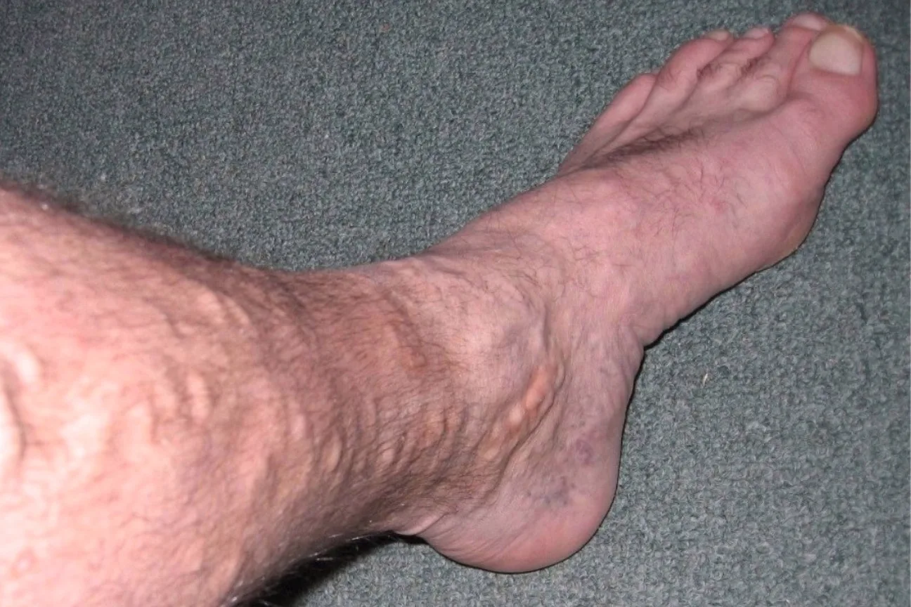
Brown staining and itchy, scaly skin around the ankles
Long-standing pressure makes the smallest vessels leak, and the pigment left behind, hemosiderin, stains the skin reddish-brown, typically around the ankles. The skin can also become itchy, swollen, dry and flaky, or scaly, crusty or blistered: stasis dermatitis, also called varicose or venous eczema. On white skin it looks red or brown; on brown or black skin it tends to look dark brown, purple or gray. Small white scars, called atrophie blanche, can appear in the same area. Stasis dermatitis explained.
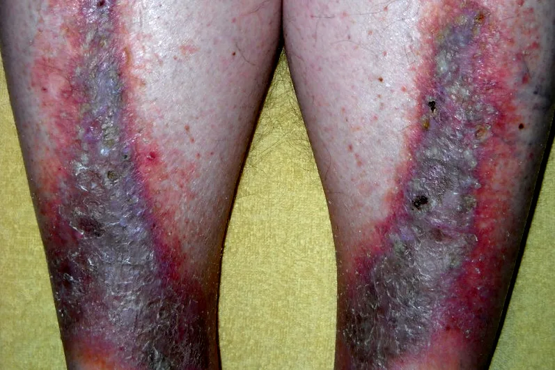
Hard, tight skin that narrows the lower leg
Over time, the skin and the fat layer beneath it on the lower leg can become tender and tight, then hard and leathery: lipodermatosclerosis. It can affect one leg or, more often, both, and the hardened lower leg can narrow, giving what DermNet describes as an upside-down champagne bottle appearance. In its early, inflamed phase it is red and painful, and can be mistaken for cellulitis.
An open sore near the ankle
Venous ulcers usually form on the inside of the lower leg, most often just above the ankle. They tend to be shallow, with uneven edges and a red base, sometimes covered with yellow tissue, and they can weep or ooze. The surrounding skin is often discolored or hard. A sore on the foot or lower leg that has not closed, or is getting bigger, wetter or more painful, should be seen the same day or the next. The NHS calls a sore that takes more than 2 weeks to heal a leg ulcer. Venous leg ulcers.
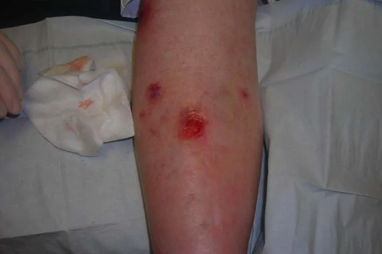
Lymph vessels: signs that fluid is not draining
In lymphedema, the lymph vessels cannot drain fluid from the tissues properly. At first the swelling is soft and leaves a dent when pressed, may come and go, and can build during the day and go down overnight, much like vein swelling. Without treatment it usually becomes more persistent, stops pitting, and the skin can harden, thicken and fold, grow wart-like bumps or leak fluid.
Two clues on the foot point toward lymphedema rather than veins: swelling that takes in the top of the foot, with toes that look squared off, and the Stemmer sign, in which a fold of skin at the base of the second toe cannot be pinched up. Repeated skin infections are common, so a swollen leg that turns red, hot and painful needs same-day care. Lymphedema: assessment and management.
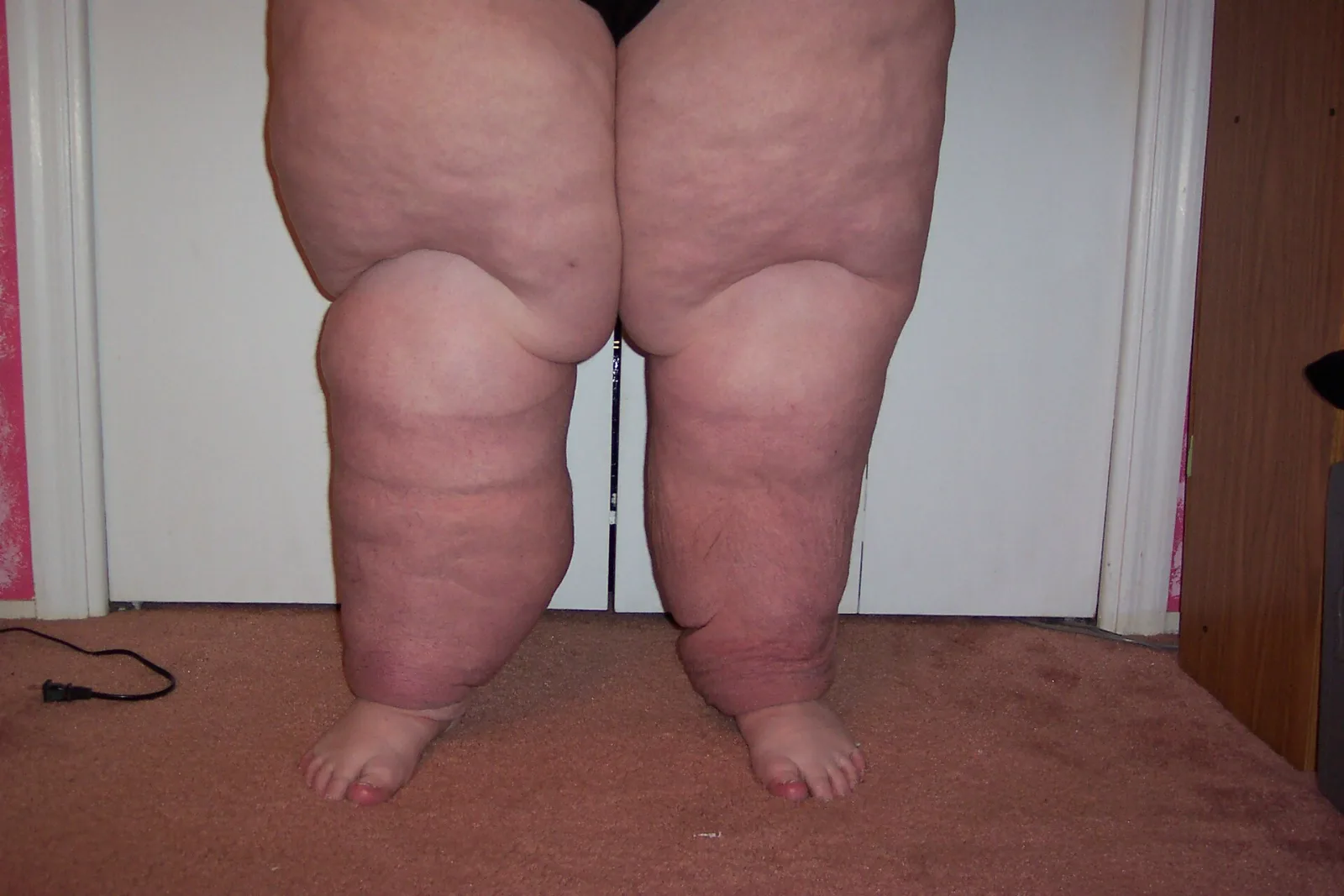
Small vessels: color changes in the cold
Raynaud’s: toes that turn white, then blue or red
Cold, anxiety or stress can trigger an attack in which the toes or fingers turn white or paler as blood flow is restricted, then may turn blue or red, often with numbness, pins and needles or pain. Attacks last from a few minutes to a few hours, and on brown or black skin the change may show mainly as paleness. See a doctor if it affects one side only, starts after age 30, or comes with joint pain, rashes or muscle weakness. Small pits or sores on a toe or fingertip that do not heal need prompt attention, and a toe sore that has not closed should be seen the same day or the next. Raynaud’s syndrome.
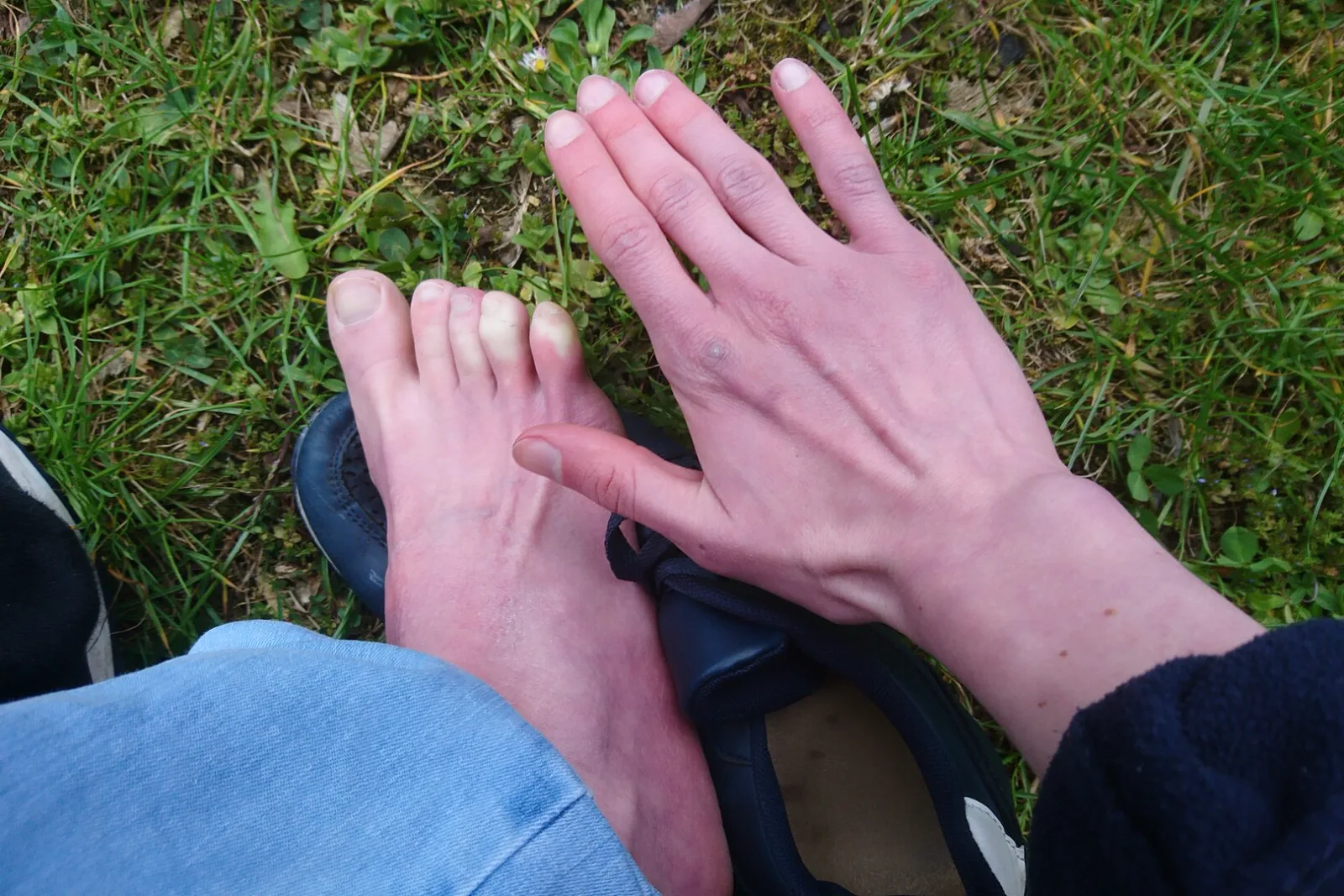
Chilblains
Small, itchy or burning red or purple patches on the toes or fingers that appear a few hours after being in the cold, as narrowed vessels near the skin widen again on warming. They usually settle in 2 to 3 weeks; see a doctor if they do not, keep coming back, ooze pus, come with a fever, or you have diabetes.
Mottling: a net-like purple pattern
A lacy, blotchy, reddish-blue to purple pattern on the legs is called livedo reticularis. In the cold it is usually a harmless reaction that fades as the skin rewarms. A pattern that stays, looks broken and irregular, or comes with sores can be linked to conditions such as antiphospholipid syndrome or vasculitis and needs a doctor’s assessment. Mottling that appears suddenly with a cold, pale, painful leg is an emergency.
Early signs: what tends to show up first
- Arteries: often nothing you can see. The usual first clue is leg pain or tiredness on walking that eases with rest, and many people notice no symptoms, so a normal-looking leg does not rule out PAD.
- Veins: heavy, tired legs and ankle swelling by evening that eases with the legs up, before any change in the skin.
- Lymph vessels: an aching, heavy limb with soft swelling that dents when pressed and goes down overnight, and clothes that fit less easily.
- Small vessels: toes that change color on cold days and recover as they warm.
An early sign that persists is worth showing to a clinician rather than watching to see whether it gets worse.
What a clinician will check
Expect an examination of your legs and feet: pulses, skin, swelling and any sores. For the arteries, the usual first test is the ankle-brachial index (ABI), which compares blood pressure at the ankle with the arm; a resting result below 0.90 suggests PAD, and a big-toe pressure is used when the ankle arteries cannot give a reliable reading. For the veins, an ultrasound shows which parts are damaged. ABI and TBI tests explained.
The order matters. Many people with vein disease also have PAD, which changes whether and how compression can be used. Anyone with diabetes, PAD symptoms, numbness, a wound, kidney disease or heart failure should have their arteries checked, usually with an ABI, before starting compression stockings. Compression safety.
Common questions
What are the early signs of poor circulation in legs?
It depends on the cause. With narrowed arteries there is often nothing to see; the usual first clue is leg pain on walking that eases with rest, and many people have no symptoms. With leaky veins, it is heavy, aching legs and ankle swelling that build through the day. Early lymphedema is soft swelling that may go down overnight.
What does poor circulation in the feet look like?
With narrowed arteries: a foot cooler or paler than the other, shiny skin with little hair, thick, slow-growing toenails, and sores on the toes or heel that are slow to heal; in advanced disease, a foot that turns red when it hangs down. Swelling of the top of the foot with squared-off toes points to lymphedema, and toes that turn white in the cold to Raynaud’s.
What are the skin signs of poor circulation?
Arterial disease tends to leave the skin shiny, tight, hairless and cool. Vein disease changes the skin around the ankles: reddish-brown staining, itchy, dry or scaly patches, then hard, tight skin and sometimes shallow sores. Lymphedema thickens the skin and can cause deep folds or wart-like growths.
What color is a leg with poor circulation?
It depends on the system. Narrowed arteries make the skin pale or bluish, and in advanced disease red when the leg hangs down. Vein disease stains the skin around the ankles reddish-brown, or dark brown, purple or gray on darker skin. Raynaud’s turns toes white, then blue or red. Sudden paleness or mottling with pain and coldness is an emergency.
How can I check my circulation at home?
You can look, but not measure. Compare both feet in the same light for temperature, color, hair, nails and sores; notice whether walking brings on calf pain that eases with rest; see whether swelling goes down overnight; and try to pinch up the skin at the base of your second toe. Only tests such as an ABI or an ultrasound can confirm a problem.
Are varicose veins a sign of poor circulation?
They are a sign of a blood-flow problem in the veins that can get worse over time, though on their own they are common and usually not serious. Aching, swelling, skin changes or a sore alongside them suggest chronic venous insufficiency, and a varicose vein that bleeds should be seen today, with a 911 call if the bleeding is heavy or does not stop with direct pressure.
Where to go next
- Poor circulation in legs and feet: causes and when to worry
- PAD symptoms and warning signs
- Chronic venous insufficiency explained
- Lymphedema: assessment and daily management
- Chilblains on toes: causes and treatment
- ABI and TBI tests: what results mean
- Near Howell or Bloomfield Township, Michigan: a foot and ankle evaluation (the author’s own practice)
Sources
- National Heart, Lung, and Blood Institute. Peripheral Artery Disease – Symptoms and Diagnosis.
- Society for Vascular Surgery. Peripheral Artery Disease, Acute Limb Ischemia and Chronic Venous Insufficiency.
- MedlinePlus. Peripheral artery disease – legs, Venous ulcers – self-care and Bleeding.
- American Academy of Family Physicians. Lower Extremity Peripheral Artery Disease: Diagnosis and Treatment (2019), and Edema: Diagnosis and Management (2013), American Family Physician.
- StatPearls, NCBI Bookshelf. Lymphedema.
- National Institute of Diabetes and Digestive and Kidney Diseases. Diabetes and Foot Problems.
- Cleveland Clinic. Chronic Venous Insufficiency (CVI), Varicose Veins and 7 Reasons Why Your Hands and Feet Are Always Cold.
- NHS. Peripheral arterial disease (PAD), Deep vein thrombosis (DVT) and Cellulitis.
- NHS. Varicose veins, Varicose eczema and Leg ulcer.
- NHS. Lymphoedema, Raynaud’s and Chilblains.
- Centers for Disease Control and Prevention. About Venous Thromboembolism (Blood Clots).
- DermNet. Lipodermatosclerosis, Livedo reticularis, Arterial ulcer and Leg ulcers.
Written by Tom Biernacki, DPM, FACFAS. Published 18 October 2026. General education, not medical advice.