Hemosiderin staining is brown, rust-colored or purple-gray discoloration of the skin, usually on the lower legs just above the inner ankles. It forms when red blood cells leak out of small veins that are under too much pressure and break down in the skin, leaving iron behind; early discoloration can also be partly the skin’s own pigment, melanin. Most often it is a sign of chronic venous insufficiency, where the leg veins struggle to send blood back up toward the heart, and doctors count it among the skin changes of advanced vein disease. The stain itself is not dangerous, but it is a good reason to have your veins checked, because the skin it sits in is more likely to break down into a leg ulcer. This guide explains what the staining is, what else can look like it, what it says about your veins, whether it fades, and what treatment can and cannot do.
When leg discoloration needs prompt care
- A sore or break in the skin that has not healed in 2 weeks: get prompt medical care, not a routine wait; leg ulcers that start over damaged veins heal faster with early treatment. If the sore is red, hot, swollen, draining pus or spreading, get same-day care, and see the next point for when to call 911. If you have diabetes, numb feet or poor circulation, get any new sore checked the same day rather than waiting 2 weeks.
- Skin that becomes painful, hot and swollen: get same-day care, because this can be cellulitis, a skin infection. Go to an emergency department now if it is spreading fast, red streaks or blood-filled blisters appear, or you have a fever, chills, shivering or flu-like symptoms, and call 911 if you also have a fast heartbeat or fast breathing, purple patches, cold, clammy or pale skin, dizziness or confusion. Cellulitis of the legs and feet →
- One leg that becomes swollen, with throbbing pain or red, blue or darker skin: get same-day care and an ultrasound that day, because a blood clot (deep vein thrombosis) has to be ruled out. Call 911 if you are also short of breath, have chest pain, cough up blood, have a fast or irregular heartbeat, or feel lightheaded or faint.
- A spot in or near the stained skin that is new, different from the others, changing, itching or bleeding: see a dermatologist promptly. Skin cancer, including melanoma, can be brown.
- Tiny red-purple spots that do not fade when pressed, or bruises, that appear for no clear reason: get a blood count within hours, because a blood or clotting problem or a medicine may be responsible. Go to an emergency department now if any are purple blotches bigger than a pencil tip, if they are widespread, or if they come with bleeding gums or nosebleeds, blood in the urine or stool, or while you take a blood thinner. Call 911 if the spots spread quickly, or come with a fever, shivering or feeling unwell, or if bleeding will not stop. Red dots on the legs and feet →
- Bleeding from a vein or a thin spot of skin on the lower leg: lie down, raise the leg above the level of your heart and press firmly on the spot. Call 911 if it does not stop or you feel faint. Even if it stops, get seen the same day: the thin skin over a high-pressure vein can bleed heavily, and it can happen again.
- A leg or foot that suddenly turns cold, pale, numb or very painful: call 911. A suddenly blocked artery is an emergency.
Redness, darkening and color change can be harder to see on brown or black skin, so pain, heat, swelling and how quickly things are changing matter more.
What hemosiderin staining is
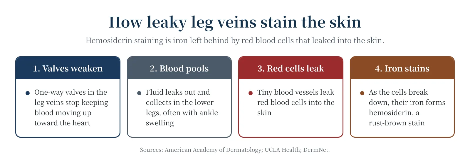
Leg veins have one-way valves that keep blood moving up toward the heart. When the valves leak, or a past clot has damaged the veins, pressure builds in the small veins of the lower leg. Fluid and red blood cells are pushed out into the skin, and the red cells break down there. As one review puts it, “The extravasation of the erythrocytes is followed by the decomposition of hemoglobin, resulting in the excessive buildup of iron, which is stored at the affected tissue as hemosiderin and causes local hyperpigmentation.” Hemosiderin is an iron-storage complex. It sits in the dermis, the deeper layer of the skin, inside the cells that cleared up the leaked blood. That is why scrubbing or exfoliating will not remove it. The iron comes from blood that leaked in that spot, so leg staining is not a sign of too much iron in your body; inherited iron overload (hemochromatosis) tends to give a more widespread bronze tint instead.
Iron is not the whole story. When researchers took small skin samples from pigmented legs, they found more melanin, the normal skin pigment, than in unstained skin, and they concluded: “Our findings suggest that in the initial phases of skin changes due to venous disease, pigmentation is attributable to melanin.” In a second study by the same team, iron deposits were found mainly in the most damaged skin: “Iron overload is not present in the less severe stages of skin damage due to CVD but lipodermatosclerosis and leg ulcers are always accompanied by haemosiderin deposition.” These are small studies from one research group, and they do not fully agree, but they suggest that early brown discoloration can be largely melanin, while iron deposits are found mainly in hardened skin and around ulcers. That difference may matter when you ask whether it will fade.
What it looks like
- Color: orange-brown, rust or reddish-brown speckles or patches on lighter skin, sometimes purple-gray. The NHS notes that the related venous eczema can look different: “On black or brown skin it tends to look dark brown, purple or grey and can sometimes be more difficult to see.”
- Place: usually just above the ankles, the area a short sock or gaiter would cover, and most often on the inner side. It is usually on both legs; on one leg it tends to point to a vein problem in that leg alone, such as an old clot.
- Company: it often comes with varicose veins, ankle swelling that is worse in the evening, a fan of small veins below the inner ankle, and dry, itchy, scaly skin (stasis dermatitis). Later the skin can become hard and tight (lipodermatosclerosis), and small white scars can appear (atrophie blanche).
Hemosiderin is also a word on brain scan and pathology reports, where it means old bleeding seen on a scan or under the microscope. This guide is about the skin of the legs only.
What causes it
Chronic venous insufficiency (the usual cause)
Most hemosiderin staining on the lower legs comes from long-standing high pressure in the leg veins. Doctors grade vein disease with the international CEAP system: in its 2020 version, pigmentation or eczema is class C4a, hardened skin or white scars C4b, and a fan of small veins at the inner ankle (corona phlebectatica) C4c. A healed ulcer is C5 and an open one C6. A systematic review of population studies, cited in the European Society for Vascular Surgery guideline, put the share of adults with C4 skin changes at about 4%. Varicose veins, a past blood clot, standing for long periods, excess weight, older age and a family history all make venous insufficiency more likely. Chronic venous insufficiency explained →
After a blood clot (post-thrombotic syndrome)
A deep vein thrombosis can scar the vein valves it passes through. Post-thrombotic syndrome, the lasting vein damage that follows, “develops in 20–50% of patients after a proximal DVT”, meaning a clot in the thigh or behind the knee. The CDC describes it this way: “People with PTS have symptoms such as swelling, pain, discoloration, and in severe cases, scaling or ulcers in the affected part of the body.” Staining on one leg only, in someone who has had a clot in that leg, often has this cause. Post-thrombotic syndrome →
After vein treatment
Brown lines or spots along treated veins are a common side effect of sclerotherapy, the injections used for spider veins and small varicose veins. In the placebo-controlled trial behind the US label for the sclerosant Asclera (polidocanol), discoloration at the injection sites occurred in 38% of people treated with Asclera, 74% with 1% sodium tetradecyl sulfate and 4% with placebo injections. Most of it fades (see below), but not all: the Sotradecol label warns, “A permanent discoloration may remain along the path of the sclerosed vein segment.” Staining also follows other vein procedures less often; the European guideline lists rates of about 1.6% to 3% after vein glue, 5% after mechanochemical ablation and 4.6% after phlebectomy. Veins lying very close to the skin carry a higher risk. Spider veins and sclerotherapy →
Bruises and injuries
Any bruise leaves hemosiderin behind as it heals, which is why bruises turn brown and yellow before they fade. On thin, sun-damaged skin, or in people taking blood thinners or steroid medicines, minor knocks can cause repeated purple bruises that leave persistent brown marks. Skin over a fracture or other injury can also stay discolored for weeks to months, occasionally more than a year, and sometimes the mark is permanent.
Schamberg’s disease and other pigmented purpuras
Pigmented purpuric dermatoses are a group of harmless conditions in which tiny skin capillaries leak. They cause clusters of pinpoint red-brown dots, often described as looking like cayenne pepper, and golden-brown patches, most often on the shins. As DermNet explains, the leaked red cells “fade away depositing haemosiderin in the upper parts of the dermis.” Schamberg’s disease is the commonest type and is more common in men. It is not a sign of vein disease and usually occurs in otherwise healthy people, though the two can coexist. A review of stasis dermatitis notes the difference under the microscope: “Hemosiderin deposition is found in the superficial dermis in PPDs, with the deposition being deeper in SD” (PPD meaning pigmented purpuric dermatosis and SD stasis dermatitis). One variety, exercise-induced capillaritis, appears on the ankles after long walks or golf in hot weather.
Minocycline and other medicines
Long-term use of minocycline, an antibiotic often taken for acne or rosacea, can stain the shins. A case report describes the pattern: “Type 2 is blue-grey pigmentation occurring in normal skin frequently on the lower extremities.” The pigment contains iron and can look like venous staining. In a 2026 case from a US vein center, a man’s legs kept darkening after successful vein treatment; a biopsy showed drug pigment after 10 years of minocycline, and stopping the drug brought partial improvement. Some other medicines, including antimalarials and amiodarone, can also discolor the skin. If your shins are blue-gray, or the color does not behave like venous staining, tell your clinician what you take.
Hemosiderin staining or something else?
Several other conditions color the lower legs. These are the ones most often confused with venous staining.
| Condition | What it looks like | Usual course |
|---|---|---|
| Venous (stasis) staining | Rust-brown to purple-gray above the inner ankles, often both legs, with swelling, varicose veins, itch or hardening | Often persists; may lighten once the veins are treated |
| Schamberg’s disease (pigmented purpura) | Pinpoint red-brown “cayenne pepper” dots and golden-brown patches on the shins; no swelling | Weeks to years; may come and go |
| Bruise or injury | Purple, then brown and yellow, at the site of a knock | Weeks; sometimes a brown mark stays longer |
| Bruising from thin skin, steroids or blood thinners | Repeated purple bruises after minor knocks, more on the forearms than the legs | 1 to 3 weeks each, sometimes leaving brown marks |
| Minocycline pigmentation | Blue-gray patches on the shins and forearms after long use | Months to years after stopping |
| Diabetic dermopathy (shin spots) | Small, round, light brown or reddish, slightly sunken spots on both shins | Usually fades over a few years |
| Darkening after a rash, scratch or burn | A brown patch exactly where the skin was inflamed or injured | Temporary; takes longer in darker skin |
| Erythema ab igne (heat marks) | A net-like red-brown pattern where a heater, hot water bottle or heating pad was used again and again | Months; severe cases may last years |
| Melanoma or another skin cancer | A single spot that is uneven in shape or color, growing, changing, itching or bleeding | Needs a dermatologist promptly |
Diabetic dermopathy deserves a mention because it is common: DermNet says the spots “usually do not require any treatment and tend to go away after a few years, particularly following improved blood glucose control.” Spots like these in someone not known to have diabetes are a reason to ask for a blood sugar test. Heat marks from a heating pad are also a warning that the skin is getting too much direct heat, which is risky for feet and legs with poor feeling or circulation.
What the staining says about your veins
On the lower legs, true venous staining means the veins have been under high pressure long enough to change the skin. National guidance treats that as a reason to look more closely. In England, NICE advises referring people with lower-leg “skin changes, such as pigmentation or eczema, thought to be caused by chronic venous insufficiency” to a vascular service. US and European vascular guidelines name a duplex ultrasound scan of the leg veins as the first test: a painless scan, usually done standing, that shows which veins are leaking and whether there is any old clot. The European guideline also recommends treating leaking surface veins in people whose vein disease has reached the skin-change stage (C4 to C6).
Will it turn into an ulcer?
Not necessarily, and nobody can give you a reliable percentage. What the research shows:
- Vein disease tends to progress. In the Edinburgh Vein Study, which re-examined 334 people with varicose veins or venous insufficiency after an average of 13.4 years, 57.8% had worsened. Nearly a third of those who started with varicose veins alone had developed venous insufficiency, and almost everyone with both had got worse. A past clot and excess weight raised the odds.
- Skin changes are the strongest warning sign. In a study comparing 120 people with varicose veins and a current or past venous ulcer with 120 people with varicose veins and no ulcer, “An increased risk of ulceration was associated with the severity of clinical venous disease, especially with the presence of skin changes (P < .0001).” Hardened skin, a fan of ankle veins and eczema each raised the odds, as did higher body weight and leaking veins behind the knee; good ankle movement and a strong calf pump protected against ulcers. A 2015 review of research on how vein disease progresses gives the same list of risk factors: “skin changes, corona phlebectatica, higher body mass index and popliteal vein reflux”.
- Staining on its own has not been studied separately. The European guideline is frank: “There is no strong evidence to guide which patients/limbs with C2 disease will progress to C4 disease, or which limbs with C4 disease are at the highest risk of progressing to ulceration.”
In practice, the risk is higher when staining comes with hardening, eczema, a past clot, excess weight or stiff ankles, and those combinations deserve attention. The measures most likely to help, such as compression, walking, ankle exercises and treating leaking veins, also ease the legs, although none has been proven to prevent a first ulcer.
Skin changes can be easier to miss on darker skin. A 2026 review of 22 studies, most of which had to use ethnicity as a stand-in for skin tone, suggested that people with darker skin can have vein disease without some of the usual visible signs, and that visible varicose veins are not a reliable marker on their own; the authors think this may delay care for people with dark skin tones. If your legs are aching, swelling or itchy, ask about your veins even if the color change is subtle.
Is hemosiderin staining dangerous?
The pigment itself is not poisonous, contagious or cancerous. The concern is the skin it sits in and what it signals. Stained skin over damaged veins is often dry and fragile, and a knock, a scratch or a tight shoe edge can open a wound that heals slowly. There is also some evidence that the iron is not just a bystander. In one study, skin around venous ulcers held about twice as much iron as comparison skin without ulcers, and the authors suggested that “dermal iron deposition may not be an incidental by-product of increased venous pressure, but may actively perpetuate tissue damage in venous ulcerations.” That is a hypothesis, not proof, but it fits with the staining being a marker of skin that is under strain. Venous leg ulcers →
Does hemosiderin staining go away?
It depends on the cause, how deep the pigment sits and how long it has been there.
| Cause | Typical course |
|---|---|
| A bruise | Changes color over a few weeks and fades; thin-skin bruises clear in 1 to 3 weeks but can leave a brown mark |
| An injury or fracture | Fades over weeks to months, sometimes more than a year; occasionally the mark is permanent |
| Sclerotherapy | Most fades within 6 to 24 months; a minority lasts longer, and some is permanent |
| Schamberg’s disease | Weeks to years, and it can come and go |
| Minocycline | Months to years after stopping the drug |
| Venous insufficiency | Long-standing staining often persists even after the veins are treated; early or mild discoloration may lighten. No study has measured how often it fades |
For staining after sclerotherapy there are real numbers. A 1987 paper on staining after sclerotherapy reported: “Eighty percent of patients who experience this adverse sequela will clear spontaneously within 6-24 months.” A 2023 systematic review found that after foam treatment of larger trunk veins, “Hyperpigmentation persisting for more than 1 year after foam polidocanol 1%-3% treatment for truncal veins has an incidence ranging from 8.1% to 17.5%.” Minocycline pigment is usually slower; a 2026 case report notes, “Usually, minocycline-induced hyperpigmentation persists beyond the 6-month to 2-year period of resolution expected in postsclerotherapy pigmentation.”
For venous staining, the honest answer is less hopeful. A 2023 review of stasis dermatitis states, “Cutaneous hyperpigmentation often persists even if the chronic venous insufficiency is treated”. The UK leg-health coalition Legs Matter puts it this way: “However, any staining present is unlikely to fully resolve, but may lighten and be less noticeable.” Treating the veins is still worth it: it eases symptoms, guidelines recommend it once vein disease has changed the skin, and it may stop new staining from forming.
Treatment: lowering the pressure in the veins
Because venous staining is a sign of vein disease, the main treatment is aimed at the veins, not the color.
- Get the veins and arteries checked. A duplex ultrasound scan shows which veins are leaking. Before you start compression of any strength, have your circulation checked too: your clinician will feel the foot pulses and, if they are weak, measure an ankle-brachial index (in diabetes, a toe pressure or an artery scan may be needed instead), because compression can damage skin when the leg’s arterial blood supply is poor. Also mention severe heart failure, or numb feet from diabetes, which can make compression unsafe. The ankle-brachial index test →
- Wear compression, once it is safe for you. For legs with hardened skin or white scars, the European guideline recommends below-knee compression stockings of 20 to 40 mmHg at the ankle to reduce the hardening. Compression also eases swelling and aching. No trial has shown that it lightens existing staining, and claims that it will prevent further staining have not been tested, but it treats the pressure that causes it. Who needs a check before compression →
- Raise your legs. The American Academy of Dermatology suggests raising your legs above your heart for 15 minutes once every two hours while you are awake if your doctor recommends it, and MedlinePlus suggests raising the leg 3 to 4 times a day for about 15 minutes each time.
- Keep your calf muscles working. Walking and ankle exercises pump blood out of the lower leg. Stiff ankles were linked to ulcers in the study above, so keeping the ankle moving matters.
- Lose weight if you are overweight. Excess weight raises the pressure in the leg veins and was a risk factor for worsening vein disease in the Edinburgh study.
- Consider treating the leaking veins. Closing a leaking surface vein with heat (radiofrequency or laser ablation), glue, foam or surgery is recommended by the European guideline for skin changes from vein disease, and it clearly helps venous ulcers heal. For skin changes other than ulcers, the guideline says the evidence is limited, and staining often remains afterwards. Vein procedures compared →
In the US, Medicare wants documented symptoms, and usually a trial of conservative care such as compression stockings, before it pays for a vein procedure, and many private insurers have similar rules; Aetna’s policy, for example, asks for a 3-month trial including medical-grade stockings when the main problem is pain and swelling. Medicare’s rules vary by region: its policy for Michigan (also Indiana, Iowa, Kansas, Missouri and Nebraska) asks for a 3-month trial even when the skin has changed, while some other regions allow the trial to be waived once vein disease has changed the skin. Compression socks for varicose veins, and the compression-trial rules by state →
Treating the color itself
Once the veins are under control, some people want the stain lightened for cosmetic reasons. The options have weak evidence, so be wary of anyone who promises clearance.
- Lasers and intense pulsed light (IPL). The evidence is a few short case reports and one small series, all reporting success, which is what you would expect when only successes get published. In the series, eight people with sclerotherapy staining more than a year old were treated with a Q-switched ruby laser: “There was significant (75-100%) resolution of hyperpigmentation in 58% of treated areas, 25% improvement in 33% of treated areas, and no improvement in the remaining areas.” The American Academy of Dermatology notes that “Some patients have seen their discoloration disappear completely after IPL”; a course of IPL typically involves three to four sessions. A 2023 review describes responses as often poor. Lasers can themselves cause light or dark patches, so choose a dermatologist experienced in treating pigment, and treat the veins first.
- Iron-binding injections. Deferoxamine, a drug that binds iron, was injected under the skin once a week in a small 2001 study of 36 women treated with sclerotherapy. Their staining reached 81% to 100% clearing in about 1 to 1½ months, compared with about 5 to 8½ months in comparison groups who were not given the drug. This is off-label, has been tested only after sclerotherapy, and is not a standard treatment.
- Lightening creams. Hydroquinone and similar creams act on melanin in the top layer of the skin. DermNet explains that “Hydroquinone lightens epidermal, but not dermal, pigmentation by reducing the production of new melanin”, so it cannot reach iron deposited deeper down. It might lighten an early melanin component, but some of the melanin in stasis skin also lies in the deeper layer, and these creams have not been tested on venous staining. We found no controlled trial of any cream sold for hemosiderin staining. Lightening creams can also irritate fragile skin or cause allergies, so ask your clinician first and never apply one to broken or eczema-affected skin.
- Cover-up makeup. Camouflage makeup made for skin discoloration can hide staining well, and dermatologists can recommend products. It is a reasonable choice; keep it off broken, weeping or eczema-affected skin.
Having sclerotherapy? Lowering the risk of staining
If you are having spider or small varicose veins injected, ask about these steps.
- Draining trapped blood. A firm, dark lump in a treated vein in the weeks after sclerotherapy is trapped blood. Letting it out with a small needle prick, called microthrombectomy, can reduce staining. In a randomized trial, “In veins 1 mm or smaller, microthrombectomy reduced pigmentation and improved overall clinical results.” The Varithena label agrees that removing it can “improve comfort and reduce skin staining.”
- Strength of the injected solution. Staining is more common with stronger concentrations of the sclerosant. The labels match the strength to the size of the vein, so ask whether the weakest suitable strength will be used.
- Compression afterwards. The evidence conflicts. One controlled study found that “Compression leads to a statistically significant reduction of post-sclerotherapy hyperpigmentation,” while a later randomized trial of three weeks of compression stockings after treatment of spider and reticular veins found that “The rate of pigmentation and matting was low and did not differ significantly between the two groups.” Follow your clinician’s advice.
- Your skin type. Staining is more likely in darker skin. Ask your clinician how they adjust treatment for it.
Caring for stained skin
Skin with venous staining is often dry, itchy and easily damaged. Moisturize it generously with a plain, fragrance-free emollient, use a soap substitute, and protect it from knocks and scratches. Flares of itchy, scaly eczema usually need a short course of a prescription steroid cream. Some ingredients often cause allergies on this skin; MedlinePlus advises asking before using creams and avoiding topical antibiotics such as neomycin, drying lotions such as calamine, lanolin, and numbing creams such as benzocaine. Stasis dermatitis: itching, discoloration and skin changes →
When to see a doctor or podiatrist
Book an appointment if:
- brown or purple staining has appeared on your lower legs and your veins have never been checked
- the stained skin is spreading, itchy, scaly or weeping, or getting hard and tight
- you have aching, heavy or swollen legs, varicose veins or a past blood clot along with the staining
- the staining is on one leg only, or does not look like the pattern described above
- a sore has opened in the stained skin, even a small one
- you take minocycline or another medicine that can discolor the skin, and the color is blue-gray
- your calves or legs ache when you walk and ease after a few minutes’ rest, your legs or feet look paler or bluer than usual, or a sore on your foot or leg is slow to heal: these can be signs of narrowed arteries (peripheral arterial disease), which also affects whether compression is safe
A vein specialist (often a vascular surgeon or interventional radiologist) can arrange a duplex scan and treat leaking veins. A dermatologist can diagnose skin conditions and treat eczema and pigment, and can take a small skin sample when the cause is unclear. A podiatrist can check the circulation, nerves and skin of the feet and ankles, look after wounds, and advise on compression that is safe for you. Mention any blood thinners, steroid medicines or minocycline, and any past clots.
Near Howell or Bloomfield Township, Michigan?
The author of this site practices podiatry at Balance Foot & Ankle, which he owns, so this is a disclosed option rather than an independent referral. If you have staining, swelling or hardening of the skin around your ankles, or a sore that is slow to heal, an evaluation can check the circulation and skin of your feet and lower legs, advise on safe compression, and coordinate a vein scan or referral to a vein specialist. Michigan evaluation →
Common questions
What is hemosiderin staining?
It is brown, rust-colored or purple-gray discoloration of the skin, usually above the ankles, caused by red blood cells that leaked out of small veins and broke down in the skin, leaving iron (hemosiderin) behind. Early discoloration can also be partly melanin, the skin’s own pigment. On the lower legs it is most often a sign of chronic venous insufficiency.
Is hemosiderin staining dangerous?
The pigment itself is not dangerous, contagious or cancerous. But staining from vein disease is a sign that the skin is under strain, and skin changes are the strongest warning sign for leg ulcers. It is worth a vein check, especially with swelling, hardening, eczema or a past clot. A single spot that is new, changing, itching or bleeding should be seen by a dermatologist.
Does hemosiderin staining go away?
Bruises and most staining after sclerotherapy fade, usually within months to two years. Long-standing staining from vein disease often persists even after the veins are treated, though it may lighten and stop spreading. Early, mild discoloration may have a better chance of fading than dark staining in hardened skin, though no study has measured this.
What is the best cream for hemosiderin staining?
No cream has been shown to remove it. Lightening creams such as hydroquinone work on melanin near the surface and cannot reach iron deposited deeper in the skin, and we found no controlled trial of any cream sold for hemosiderin staining. Lightening creams can also irritate fragile skin or cause allergies, so ask your clinician first and never apply one to broken or eczema-affected skin. Plain moisturizers are still worth using, because they protect fragile skin, and camouflage makeup can hide the color.
Can laser treatment remove hemosiderin staining?
Sometimes it lightens it. Short reports describe improvement with several types of laser and with intense pulsed light, but they are case reports and one small series, and one review describes responses as often poor. Treat the leaking veins first, choose a dermatologist experienced in treating pigment, and expect several sessions and a chance of new light or dark patches.
What does early hemosiderin staining look like?
Usually small reddish-brown or rust-colored speckles or a faint brown patch just above the inner ankle, often on both legs, sometimes with mild ankle swelling, itch or varicose veins. On brown or black skin it may look darker brown, purple or gray and be harder to see. This early stage is the best time to have the veins checked.
Do compression stockings help hemosiderin staining?
They treat the high vein pressure that causes it, ease swelling and aching, and reduce skin hardening, but no study has shown that they lighten existing stains. Have your circulation checked before starting compression of any strength, even over-the-counter socks, and get the right size and strength. Compression socks for vein disease →
What is the difference between hemosiderin staining and Schamberg’s disease?
Both leave hemosiderin in the skin, but for different reasons. Schamberg’s disease is a harmless leak from tiny capillaries that causes pinpoint “cayenne pepper” dots and golden-brown patches, usually on the shins, in otherwise healthy people. Venous staining comes from high pressure in the leg veins, sits deeper in the skin, and usually comes with swelling, varicose veins or eczema around the ankles.
Is hemosiderin staining the same as stasis dermatitis?
They are related but not the same. Stasis dermatitis is the itchy, dry, scaly inflammation of the skin that venous insufficiency causes; hemosiderin staining is the brown discoloration that often comes with it. Both are skin changes of vein disease. The eczema can usually be calmed with skin care, steroid creams and treatment of the vein pressure, but the brown staining often remains. Stasis dermatitis →
Where to go next
- Stasis dermatitis: itching, discoloration and skin changes
- Chronic venous insufficiency and venous stasis
- Varicose veins: symptoms and treatment choices
- Spider veins on the legs: causes, when to worry and treatment
- Venous leg ulcers: why compression is the treatment
- Compression safety: who needs a check first
Sources
- Silverberg J, Jackson JM, Kirsner RS, et al. Narrative review of the pathogenesis of stasis dermatitis: an inflammatory skin manifestation of venous hypertension. Dermatology and Therapy 2023;13:935–950.
- Caggiati A, Rosi C, Franceschini M, Innocenzi D. The nature of skin pigmentations in chronic venous insufficiency: a preliminary report. European Journal of Vascular and Endovascular Surgery 2008;35:111–118.
- Caggiati A, Rosi C, Casini A, et al. Skin iron deposition characterises lipodermatosclerosis and leg ulcer. European Journal of Vascular and Endovascular Surgery 2010;40:777–782.
- Kim D, Kang WH. Role of dermal melanocytes in cutaneous pigmentation of stasis dermatitis: a histopathological study of 20 cases. Journal of Korean Medical Science 2002;17:648–654.
- Yosipovitch G, Nedorost ST, Silverberg JI, et al. Stasis dermatitis: an overview of its clinical presentation, pathogenesis, and management. American Journal of Clinical Dermatology 2023;24:275–286.
- NHS. Varicose eczema; Leg ulcer; Cellulitis; Deep vein thrombosis (DVT); Peripheral arterial disease (PAD); Sepsis.
- DermNet. Venous eczema; Venous insufficiency; Lipodermatosclerosis; Capillaritis (pigmented purpura); Purpura; Senile purpura; Drug-induced skin pigmentation; Diabetic skin disease; Postinflammatory hyperpigmentation; Erythema ab igne; Hydroquinone.
- De Maeseneer MG, Kakkos SK, Aherne T, et al. European Society for Vascular Surgery (ESVS) 2022 clinical practice guidelines on the management of chronic venous disease of the lower limbs. European Journal of Vascular and Endovascular Surgery 2022;63:184–267.
- Lurie F, Passman M, Meisner M, et al. The 2020 update of the CEAP classification system and reporting standards. Journal of Vascular Surgery: Venous and Lymphatic Disorders 2020;8:342–352.
- Gloviczki P, Lawrence PF, Wasan SM, et al. The 2023 Society for Vascular Surgery, American Venous Forum, and American Vein and Lymphatic Society clinical practice guidelines for the management of varicose veins of the lower extremities. Part II. Journal of Vascular Surgery: Venous and Lymphatic Disorders 2024;12:101670.
- National Institute for Health and Care Excellence. Varicose veins: diagnosis and management (CG168), recommendations.
- Makedonov I, Kahn SR, Galanaud JP. Prevention and management of the post-thrombotic syndrome. Journal of Clinical Medicine 2020;9:923.
- Centers for Disease Control and Prevention. About venous thromboembolism (blood clots).
- DailyMed, US National Library of Medicine. Asclera (polidocanol) injection label; Sotradecol (sodium tetradecyl sulfate) injection label; Varithena (polidocanol injectable foam) label.
- Schadler ED, Cibull TL, Mehlis SL. A severe case of minocycline-induced hyperpigmentation of the lower extremities. Cureus 2018;10:e2672.
- Doshi M, Sak L, Jimenez JC. Drug-induced hyperpigmentation confounding clinical assessment and management for advanced chronic venous insufficiency. Journal of Vascular Surgery Cases, Innovations and Techniques 2026;12:102327.
- Tolaymat L, Hall MR. Pigmented purpuric dermatosis; Zaldivar Fujigaki JL, Anjum F. Schamberg disease; Hoskin J, Kaneko K, Munakomi S, Fife T. Superficial siderosis. StatPearls.
- Lee AJ, Robertson LA, Boghossian SM, et al. Progression of varicose veins and chronic venous insufficiency in the general population in the Edinburgh Vein Study. Journal of Vascular Surgery: Venous and Lymphatic Disorders 2015;3:18–26.
- Robertson L, Lee AJ, Gallagher K, et al. Risk factors for chronic ulceration in patients with varicose veins: a case control study. Journal of Vascular Surgery 2009;49:1490–1498.
- Pannier F, Rabe E. Progression in venous pathology. Phlebology 2015;30(1 Suppl):95–97.
- Clemett VJ, Geraghty J, Oozageer Gunowa N, et al. A systematic review of the effectiveness of assessing skin changes for chronic venous insufficiency in people with dark skin tones and the impact on the patient journey and clinical care. International Journal of Vascular Medicine 2026;2026:8034303.
- Ackerman Z, Seidenbaum M, Loewenthal E, Rubinow A. Overload of iron in the skin of patients with varicose ulcers: possible contributing role of iron accumulation in progression of the disease. Archives of Dermatology 1988;124:1376–1378.
- Goldman MP, Kaplan RP, Duffy DM. Postsclerotherapy hyperpigmentation: a histologic evaluation. Journal of Dermatologic Surgery and Oncology 1987;13:547–550.
- Bossart S, Daneluzzi C, Cazzaniga S, et al. Skin hyperpigmentation after sclerotherapy with polidocanol: a systematic review. Journal of the European Academy of Dermatology and Venereology 2023;37:274–283.
- Legs Matter. Haemosiderin.
- American Academy of Dermatology. Stasis dermatitis: diagnosis and treatment; What to look for: ABCDEs of melanoma.
- MedlinePlus, US National Library of Medicine. Stasis dermatitis and ulcers; Bleeding into the skin.
- Tafazzoli A, Rostan EF, Goldman MP. Q-switched ruby laser treatment for postsclerotherapy hyperpigmentation. Dermatologic Surgery 2000;26:653–656.
- Laser and light reports: Wu DC, Goldman MP. Successful treatment of chronic venous stasis hyperpigmentation of the lower limbs with the picosecond alexandrite laser. Dermatologic Surgery 2018;44:881–883; Pimentel CL, Rodriguez-Salido MJ. Pigmentation due to stasis dermatitis treated successfully with a noncoherent intense pulsed light source. Dermatologic Surgery 2008;34:950–951; Hamilton HK, Dover JS, Arndt KA. Successful treatment of disfiguring hemosiderin-containing hyperpigmentation with the Q-switched 650-nm wavelength laser. JAMA Dermatology 2014;150:1221–1222; Gan SD, Orringer JS. Hemosiderin hyperpigmentation: successful treatment with Q-switched 755-nm laser therapy. Dermatologic Surgery 2015;41:1443–1444; Swali RN, Rajanala S, Friedman PM. Treatment of hemosiderin staining with a 785-nm picosecond titanium sapphire laser in combination with a nonablative 1550-nm fractional resurfacing laser. Dermatologic Surgery 2025;51:108–109.
- Lopez L, Dilley RB, Henriquez JA. Cutaneous hyperpigmentation following venous sclerotherapy treated with deferoxamine mesylate. Dermatologic Surgery 2001;27:795–798.
- Scultetus AH, Villavicencio JL, Kao TC, et al. Microthrombectomy reduces postsclerotherapy pigmentation: multicenter randomized trial. Journal of Vascular Surgery 2003;38:896–903.
- Weiss RA, Sadick NS, Goldman MP, Weiss MA. Post-sclerotherapy compression: controlled comparative study of duration of compression and its effects on clinical outcome. Dermatologic Surgery 1999;25:105–108; Kern P, Ramelet AA, Wütschert R, Hayoz D. Compression after sclerotherapy for telangiectasias and reticular leg veins: a randomized controlled study. Journal of Vascular Surgery 2007;45:1212–1216.
- Centers for Medicare & Medicaid Services, Medicare Coverage Database. L34536: Treatment of varicose veins of the lower extremities (WPS, which covers Michigan) and L34924: Treatment of chronic venous insufficiency of the lower extremities (Novitas). Aetna. Varicose veins: medical clinical policy bulletin 0050.
- Rodrigues A, Puggina J, da Silva VS, et al. Pigmentation after foam or cryo-laser cryo-sclerotherapy for lower limb reticular veins: a within-patient randomized trial. Journal of Vascular Surgery: Venous and Lymphatic Disorders 2025;13:102285.
- Rabe E, Partsch H, Morrison N, et al. Risks and contraindications of medical compression treatment: a critical reappraisal. An international consensus statement. Phlebology 2020;35:447–460.
- UCLA Health, Ask the Doctors. Hemosiderin staining product of trauma or venous insufficiency. 2 January 2023.
Written by Tom Biernacki, DPM, FACFAS. Published 12 November 2026. General education, not medical advice.