Feet change color for reasons that range from a cold kitchen floor to a blocked artery. The color itself is rarely the answer. What matters is the pattern: which foot, how fast it appeared, what position brings it on, and what else is happening to the skin.
Get help immediately for these
Call 911 if a foot or leg becomes pale, mottled or blue over minutes to hours and is also cold, numb, weak or severely painful. That combination can mean blood supply has been cut off suddenly, and permanent damage can happen within four to six hours.
Same day for new redness that is hot and tender, and for a toe that has turned dusky blue or purple but is still soft. Go to an emergency department now if redness spreads fast, leaves a red streak or comes with a fever or chills (with diabetes, numb feet or poor circulation, for any spreading redness); for a toe or patch of skin that has turned black, gray or dark purple, or looks dead; and for skin that has turned white or grayish-yellow, waxy and numb after freezing cold, which can be frostbite.
Why feet change color
Skin color at the surface reflects how much blood is in the small vessels just under it and how much oxygen that blood is carrying. Three things move it: how much blood the arteries deliver, how well the veins carry it away, and how the tiny vessels themselves respond to temperature, position and nerve signals.
That is why position matters so much. Gravity pulls blood into a dangling foot and drains it from a raised one. In a healthy leg the color barely shifts. In a leg with poor arterial supply, the difference can be dramatic — and that contrast is more informative than any single color.
The main patterns
| What you notice | What tends to come with it | What it can point to |
|---|---|---|
| Pale or white when the leg is raised | Returns slowly to color when lowered, often over 30 seconds or more | Reduced arterial supply |
| Deep red or purplish when the foot hangs down | Fades when the leg is raised; often paired with the pallor above | Advanced arterial disease. This pairing, called dependent rubor, is one of the more telling findings on the foot |
| Blue or gray-purple, both feet, cool | Settles with warming; fingers may do it too | Often normal response to cold; persistent change deserves a look (acrocyanosis is one cause) |
| White, then blue, then red on cold or stress | Sharply bordered, reverses on warming, tingling as it recovers; attacks often last around 20 minutes | Raynaud’s phenomenon |
| Itchy red or purple patches on the toes | Appear a few hours after damp cold that is not freezing; may swell; usually clear in two to three weeks | Chilblains, including the “COVID toes” pattern |
| White or grayish-yellow, firm or waxy, numb | After freezing temperatures | Frostbite. Do not rub it or rewarm it with direct heat; go to an emergency department now |
| One toe suddenly blue or purple, with pulses still present | Painful, sharply demarcated, rest of the foot looks normal | A small embolus lodging in a digital artery (blue toe syndrome) — needs same-day assessment |
| Red, hot, burning feet that flare with warmth or exercise | Eased by cooling and raising the feet; usually both feet | Erythromelalgia; cool with air, not ice water |
| Bright red, hot, swollen, tender, spreading | Fever, chills, a wound or crack in the skin | Infection (cellulitis) |
| Rusty, red-brown or tan staining around the ankle | Swelling, itching, dry flaky skin, prominent veins | Long-standing venous disease staining the skin with iron (hemosiderin staining) |
| Net-like blue-purple mottling | Comes and goes with temperature | Often harmless in cold (livedo reticularis); new, fixed or painful mottling needs assessment |
| Black, dark brown or leathery patch | Firm, often on a toe tip, heel or pressure point | Tissue that has lost its blood supply. Urgent |
| Yellow-brown thick nails, no skin change | Crumbly, slow-growing nails | Often fungal, but injury, shoe pressure and poor circulation also thicken nails; a lab test confirms fungus before treatment |
Raynaud’s, briefly
Raynaud’s affects an estimated 3 to 5% of adults worldwide, more often women. It is a spasm of the small vessels rather than a blockage, and many people only ever see one or two of the three colors. Primary Raynaud’s is much more common and usually mild; the secondary form, which comes with conditions such as scleroderma or lupus, can be more serious. The NHS advises seeing a doctor if attacks affect one side only or start for the first time after about age 30, and any sore on a toe or fingertip should be checked. Raynaud’s and cold feet in detail →
Color is harder to read on brown and black skin
Redness, pallor and blue discoloration are described in most medical writing as they appear on white skin, and they are genuinely easier to miss on darker skin. Pallor may look ashen or gray rather than white; redness may look darker, purple or simply different from the surrounding skin rather than obviously red; and early infection may show as darkening rather than reddening. The NHS notes that the skin changes of venous eczema can look dark brown, purple or gray on darker skin rather than red.
Two things help. First, compare with the same spot on the other foot and with skin further up the leg. Second, use what you can feel rather than only what you can see: warmth, swelling, firmness, tenderness and the difference between the two feet carry the same information and do not depend on color at all.
What to check yourself
- Compare the two feet in the same light. A change on one foot only is far more meaningful than both feet looking the same.
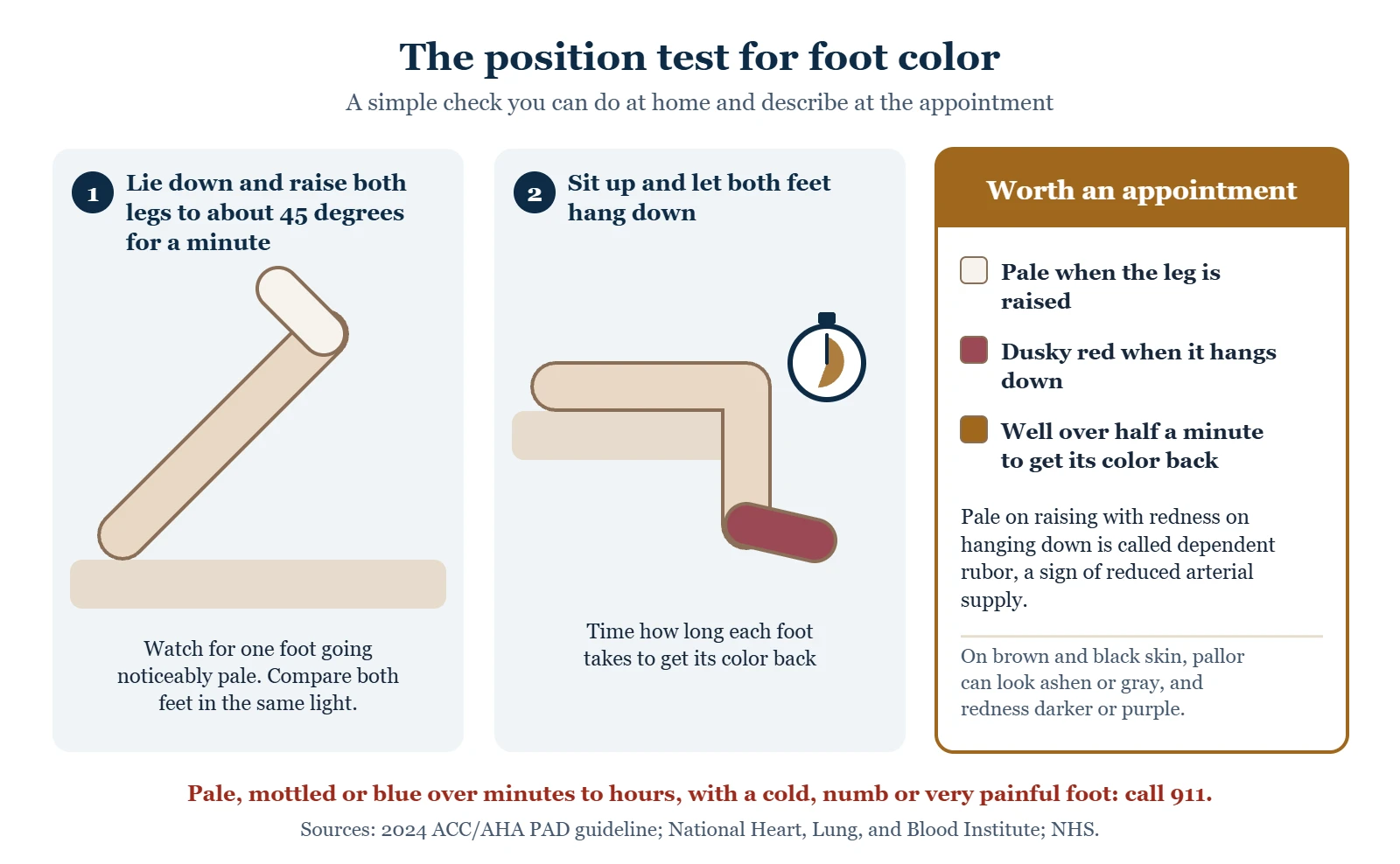
- Try the position test. Lie down and raise both legs to about 45 degrees for a minute. Note whether one foot goes noticeably pale. Then sit up and hang both feet down, and time how long each takes to return to its normal color. A foot that blanches on raising and then turns dusky red hanging down, taking well over half a minute to recolor, is worth an appointment.
- Note how fast it appeared. Minutes to hours is urgent. Weeks to months is a different conversation.
- Feel the temperature. Use the back of your hand on both feet and compare with your calves.
- Check for a break in the skin. Any wound, crack or blister on a discolored foot raises the priority.
- Look at the skin and hair. Shiny, thin, hairless skin and thickened nails travel with reduced arterial supply.
- Photograph it. Color changes come and go, and they rarely oblige by appearing in the exam room. A photo in daylight, both feet in frame, is genuinely useful to whoever sees you.
What a clinician will do
Feel the pulses in both feet and behind both knees, look at skin, hair, nails and the spaces between the toes, test sensation, and compare the two sides. If anything points at the arteries, the next step is usually an ankle-brachial index, which is quick and painless. Toe pressures are used when the ankle vessels are stiff, which is common in diabetes and kidney disease. Ultrasound follows if the picture needs more detail.
If the pattern looks venous instead, the examination focuses on swelling, skin texture and staining around the ankle, and ultrasound looks at the valves in the leg veins. If Raynaud’s looks secondary, blood tests for autoimmune disease are usually part of the workup.
What not to do
Do not apply direct heat to a discolored foot — no heating pads, hot water bottles or hot soaks. If sensation is reduced or arterial supply is poor, burns happen easily and heal badly; the CDC gives the same warning for frostbitten skin, which also should not be rubbed. Do not start compression on a foot that has changed color until someone has checked the arterial supply, because compression on a poorly supplied leg can make things worse. Why → And do not wait out a change that appeared over hours.
From the clinic
The single most useful thing a patient can bring me is a photo of the foot at its worst, next to the other foot, in daylight. Color changes rarely perform on cue in the exam room, and “it goes purple sometimes” can mean anything from a cold floor to an artery problem. Two feet side by side, and a note of what position you were in, usually tells me which direction to look.
Common questions
My feet turn purple when I sit for a while. Is that serious?
On its own, often not. Blood pools in the feet when they hang down and the small vessels relax, and in cool rooms plenty of healthy feet go blotchy purple. What changes the picture is if the color stays after you walk around or raise the legs, if only one foot does it, if the foot is also cold or painful, or if the skin is breaking down. Those deserve an appointment. Feet that turn purple when you sit: dependent acrocyanosis →
What does it mean if my foot is red when it hangs down but pale when I lift it?
That combination — pallor on elevation, dusky redness on dependency — is one of the more specific signs of significantly reduced arterial supply to the foot, and it is worth taking to a clinician rather than watching. It is not an emergency by itself if the foot is warm, comfortable and intact, but it should lead to an ankle-brachial index fairly promptly. More on PAD symptoms →
Is the brown staining around my ankles permanent?
Largely, yes. The discoloration comes from iron left behind when red cells leak out of overloaded small veins, and once it is in the skin it fades very slowly if at all. What treatment does is stop it advancing and protect the skin from breaking down, which matters more than the color. How venous insufficiency causes it →
My toe went blue overnight but my foot pulses are fine. Why?
A single blue or purple toe with normal pulses often means a small fragment has traveled from further up and lodged in one of the tiny arteries of the toe. Because the blockage is downstream of where pulses are felt, the pulses stay normal — which is exactly why this one gets dismissed. It needs assessment within a day or so, because the source upstream is the real question.
What is the lacy purple pattern on my legs?
A purple or red-blue net on the legs that appears in the cold and fades when you warm up is usually livedo reticularis in its harmless form, which is common in babies and young women. A pattern that stays when you are warm, looks broken or irregular, or comes with painful lumps or sores needs a check. Purple patches that do not fade when pressed need an emergency department now, and 911 if they appear suddenly, spread quickly, or come with a fever or feeling unwell. Livedo reticularis: causes and when to worry →
Can medication cause my feet to change color?
Yes. Cold hands or feet is a common side effect of beta blockers, and ergotamine migraine medicines and decongestants containing pseudoephedrine or phenylephrine can make cold-triggered color changes more pronounced. A few drugs cause mottling or discoloration directly. It is worth raising with whoever prescribes them rather than stopping anything on your own. Medicines and leg changes →
Are chilblains the same as frostbite?
No. Chilblains come from damp cold that is not freezing: itchy red or purple patches appear on the toes a few hours later and usually clear in two to three weeks, though they are worth getting checked if they do not improve or if you have diabetes. Frostbite is tissue actually freezing — white or grayish-yellow skin that feels firm or waxy and numb — and it needs an emergency department now: the hospital treatments that save tissue work only within hours to days.
Where to go next
- Trench foot: pale, then red and swollen, after cold and wet
- Gangrene: when a toe turns black, or wet and swollen
- Red dots on the legs and feet: petechiae and when to worry
- Frostbite: white, gray or waxy toes after freezing cold
- Cold feet: what it does and does not mean
- PAD symptoms and warning signs
- Brown, itchy skin above the ankles: stasis dermatitis
- When to seek care
- Poor circulation in the legs and feet
- Raynaud’s syndrome: fingers and toes that turn white, then blue
- Chilblains on the toes: causes, treatment and when to worry
- Signs of poor circulation in the legs and feet, with pictures
Sources
- National Heart, Lung, and Blood Institute. Peripheral Artery Disease — Symptoms.
- Society for Vascular Surgery. Acute Limb Ischemia.
- Cleveland Clinic. Acute limb ischemia.
- Gornik HL, et al. 2024 ACC/AHA multisociety guideline for the management of lower extremity peripheral artery disease. Circulation 2024.
- MedlinePlus Genetics. Raynaud phenomenon.
- Cleveland Clinic. Raynaud’s phenomenon.
- NHS. Raynaud’s.
- National Institute of Arthritis and Musculoskeletal and Skin Diseases. Raynaud’s phenomenon.
- Mayo Clinic. Chilblains.
- NHS. Chilblains.
- American Academy of Dermatology. COVID toes.
- CDC. Preventing frostbite.
- NHS. Varicose eczema.
- NHS. Leg ulcer — symptoms.
- Mayo Clinic. Beta blockers.
- Centers for Disease Control and Prevention. About Blood Clots.
- National Institute of Diabetes and Digestive and Kidney Diseases. Diabetes and Foot Problems.
Written by Tom Biernacki, DPM, FACFAS. Published 21 September 2026. Updated 15 October 2026. General education, not medical advice.